Volume 30, Number 3—March 2024
Dispatch
Taenia martis Neurocysticercosis-Like Lesion in Child, Associated with Local Source, the Netherlands
Figure 1
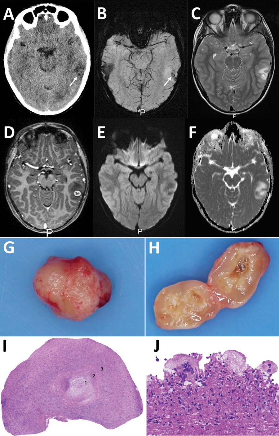
Figure 1. Diagnostic imaging of the brain and cystic lesion resected from boy with neurocysticercosis-like lesion, the Netherlands. A) Axial computed tomography showing edema in the left temporal lobe with a barely visible hypointense round lesion with a noncalcified, isointense rim (arrow). B–F) Axial magnetic resonance images at slightly different levels through the cystic lesion with surrounding edema in the left temporal lobe, showing a hypointense ring on susceptibility-weighted image (B) using minimum intensity projection (arrow) and on T2-weighted image (C), suggestive of a fibrotic capsule. D) Three-dimensional T1-weighted image showing a slightly irregular enhancement of the rim. E, F) On diffusion-weighted image (E) and apparent diffusion coefficient map (F), the rim is isointense and central diffusion restriction is absent, excluding a bacterial abscess. G, H) Macroscopic picture of the lesion showing a round nodule (G) and a cyst-like lesion (H) on cut section with a white-greyish central area surrounded by a thin capsule. I, J) Microscopic images showing a necrotic core (1) surrounded by a rim of fibrosis (2) and a mixed inflammatory response (3) (I) and multinuclear foreign-body-type giant cells (J).
1These first authors contributed equally to this article.