Skip directly to site content Skip directly to page options Skip directly to A-Z link Skip directly to A-Z link Skip directly to A-Z link
Volume 30, Number 6—June 2024
Dispatch

Encephalitozoon cuniculi Microsporidia in Cerebrospinal Fluid from Immunocompetent Patients, Czech Republic

Bohumil SakComments to Author , Katka Mansfeldová, Klára Brdíčková, Petra Gottliebová, Elka Nyčová, Nikola Holubová, Jana Fenclová, Marta Kicia, Żaneta Zajączkowska, and Martin Kváč
Author affiliations: Biology Centre of the Czech Academy of Sciences, České Budějovice, Czech Republic (B. Sak, N. Holubová, J. Fenclová, M. Kváč); University of South Bohemia, České Budějovice (K. Mansfeldová, J. Fenclová, M. Kváč); Bulovka Hospital, Prague, Czech Republic (K. Brdíčková, P. Gottliebová, E. Nyčová); Wroclaw Medical University, Wroclaw, Poland (M. Kicia, Ż. Zajączkowska)

Main Article

Figure 1

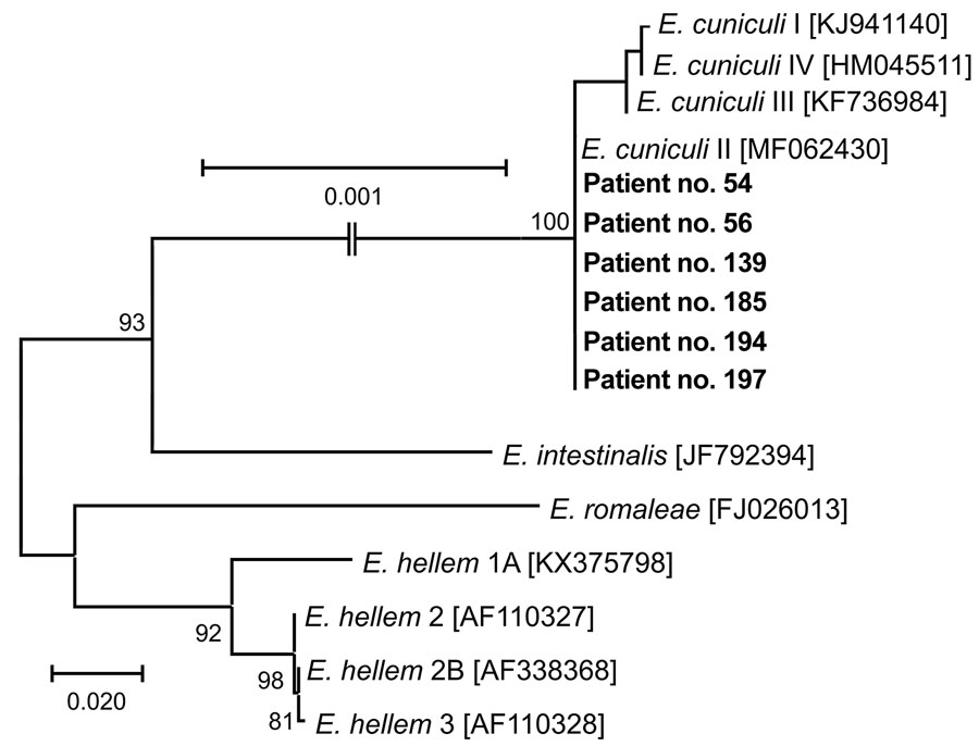
Phylogenetic analysis of Encephalitozoon cuniculi genotypes recovered from cerebrospinal fluid of immunocompetent patients, Czech Republic. Bold indicates sequences obtained in this study, identified by patient number. Sequences for comparisons were obtained from GenBank; accession numbers are in brackets. Tree was constructed by using the maximum-likelihood method. Partial sequences of 16S rRNA gene, the entire internal transcribed spacer region, and a partial sequence of 5.8S rRNA gene were inferred by using neighbor-joining analyses, and relationships were computed by using the Tamura 3-parameter method with gamma distribution and parametric bootstrap analysis of 1,000 replicates in MEGA X software (MEGA, https://www.megasoftware.net). Scale bar indicates nucleotide substitutions per site.

Figure 1. Phylogenetic analysis of Encephalitozoon cuniculi genotypes recovered from cerebrospinal fluid of immunocompetent patients, Czech Republic. Bold indicates sequences obtained in this study, identified by patient number. Sequences for comparisons were obtained from GenBank; accession numbers are in brackets. Tree was constructed by using the maximum-likelihood method. Partial sequences of 16S rRNA gene, the entire internal transcribed spacer region, and a partial sequence of 5.8S rRNA gene were inferred by using neighbor-joining analyses, and relationships were computed by using the Tamura 3-parameter method with gamma distribution and parametric bootstrap analysis of 1,000 replicates in MEGA X software (MEGA, https://www.megasoftware.net). Scale bar indicates nucleotide substitutions per site.

Main Article

Page created: April 19, 2024
Page updated: May 22, 2024
Page reviewed: May 22, 2024
The conclusions, findings, and opinions expressed by authors contributing to this journal do not necessarily reflect the official position of the U.S. Department of Health and Human Services, the Public Health Service, the Centers for Disease Control and Prevention, or the authors' affiliated institutions. Use of trade names is for identification only and does not imply endorsement by any of the groups named above.
file_external