Skip directly to site content Skip directly to page options Skip directly to A-Z link Skip directly to A-Z link Skip directly to A-Z link
Volume 30, Number 6—June 2024
Research Letter

Characterization of Cetacean Morbillivirus in Humpback Whales, Brazil

Derek B. de AmorimComments to Author , Laura J. de Camargo, Paula R. Ribeiro, Renata da F. Budaszewski, Jean Carlo O. Menegatt, Milena C. Paz, Lucas T. de Castro, Paula R. Almeida, Juliana C. Olegário, Cláudio W. Canal, and Luciana Sonne
Author affiliations: Universidade Federal do Rio Grande do Sul, Imbé, Brazil (D.B. de Amorim); Universidade Federal do Rio Grande do Sul, Porto Alegre, Brazil (L.J. Camargo, P.R. Ribeiro, R.F. Budaszewski, J.C.O. Menegatt, M.C. Paz, L.T. de Castro, J.C. Olegário, C.W. Canal, L. Sonne); Universidade FEEVALE–Campus II, Novo Hamburgo, Brazil (P.R. Almeida)

Main Article

Figure

Microscopic findings of cetacean morbillivirus infection in 2 humpback whales in southern Brazil, 2022 (Megaptera novaeangliae). A) Cerebral cortex from whale MN2,. Note the pronounced perivascular cuffs composed of lymphocytes and plasma cells, moderate gliosis, and discreet vacuolization of the white matter. Hematoxylin and eosin stain; original magnification ×200. B) Cerebral cortex from whale. Eosinophilic intracytoplasmic inclusion body in a neuronal cell (arrow). Hematoxylin and eosin stain; original magnification ×400. C) Cerebral cortex from whale MN2. Neurons and astrocytes show severe, multifocal, cytoplasmic immunostaining with a marked perivascular lymphoplasmacytic cuff. Immunohistochemistry anti-canine distemper virus, morbillivirus; original magnification ×100. D) Cerebellum from MN2. Purkinje cells exhibit pronounced, multifocal, cytoplasmic immunostaining. Inset: Intense and granular immunostaining is observed in the cell body, in the dendrites of Purkinje cells, and occasionally in granule cells. Immunohistochemistry anti-canine distemper virus, morbillivirus; original magnification ×400.

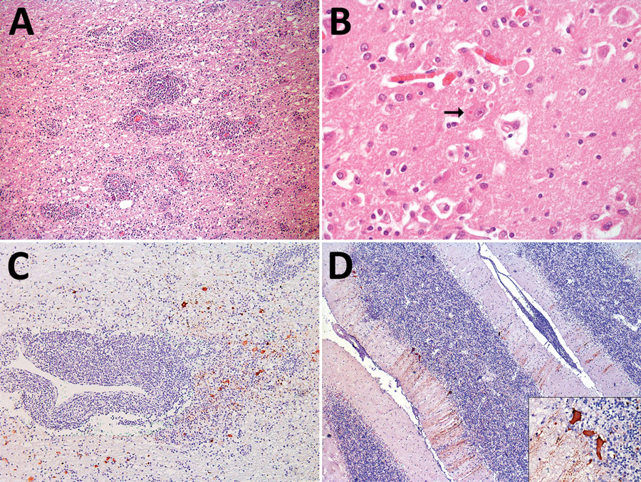
Figure. Microscopic findings of cetacean morbillivirus infection in 2 humpback whales in southern Brazil, 2022 (Megaptera novaeangliae). A) Cerebral cortex from whale MN2,. Note the pronounced perivascular cuffs composed of lymphocytes and plasma cells, moderate gliosis, and discreet vacuolization of the white matter. Hematoxylin and eosin stain; original magnification ×200. B) Cerebral cortex from whale. Eosinophilic intracytoplasmic inclusion body in a neuronal cell (arrow). Hematoxylin and eosin stain; original magnification ×400. C) Cerebral cortex from whale MN2. Neurons and astrocytes show severe, multifocal, cytoplasmic immunostaining with a marked perivascular lymphoplasmacytic cuff. Immunohistochemistry anti-canine distemper virus, morbillivirus; original magnification ×100. D) Cerebellum from MN2. Purkinje cells exhibit pronounced, multifocal, cytoplasmic immunostaining. Inset: Intense and granular immunostaining is observed in the cell body, in the dendrites of Purkinje cells, and occasionally in granule cells. Immunohistochemistry anti-canine distemper virus, morbillivirus; original magnification ×400.

Main Article

Page created: May 02, 2024
Page updated: May 22, 2024
Page reviewed: May 22, 2024
The conclusions, findings, and opinions expressed by authors contributing to this journal do not necessarily reflect the official position of the U.S. Department of Health and Human Services, the Public Health Service, the Centers for Disease Control and Prevention, or the authors' affiliated institutions. Use of trade names is for identification only and does not imply endorsement by any of the groups named above.
file_external