Rickettsia parkeri Rickettsiosis in Kidney Transplant Recipient, North Carolina, USA, 2023
Gautam M. Phadke

, Kiran Gajurel, Jennifer Kasten, Marlene DeLeon-Carnes, Carmen Ramos, Sandor E. Karpathy, Arlyn N. Gleaton, Sydney N. Adams, Pallavi D. Annambhotla, Sridhar V. Basavaraju, Carl Williams, and Christopher D. Paddock
Author affiliations: Metrolina Nephrology Associates, Charlotte, North Carolina, USA (G. Phadke); Carolinas Medical Center, Atrium Health, Charlotte (G. Phadke, K. Gajurel); Centers for Disease Control and Prevention, Atlanta, Georgia, USA (J. Kasten, M. DeLeon-Carnes, C. Ramos, S. Karpathy, A.N. Gleaton, S.N. Adams, P.D. Annambhotla, S.V. Basavaraju, C.D. Paddock); Oak Ridge Institute for Science and Education, Oak Ridge, Tennessee, USA (S.N. Adams); North Carolina Department of Health and Human Services, Raleigh, North Carolina, USA (C. Williams)
Main Article
Figure 2
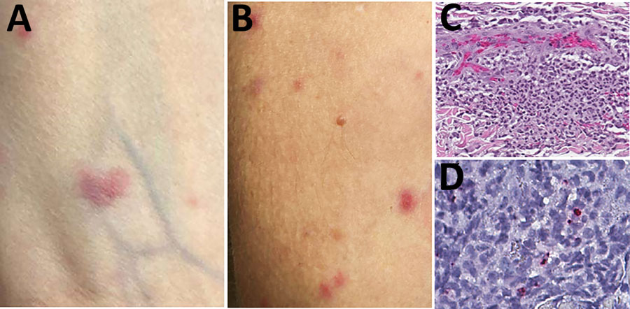
Figure 2. Skin lesions and testing results for a kidney transplant receipient diagnosed with Rickettsia parkeri rickettsiosis, North Carolina, USA, 2023. A, B) Sparse maculopapular rash involving forearms. Lesions ranged from 0.2 to 3 cm in greatest dimension and were tender and erythematous. C) Histopathologic appearance of rash lesion demonstrating perivascular collections of mixed inflammatory cell infiltrates in the mid-dermis comprising predominantly neutrophils and macrophages. Hematoxylin and eosin stain; original magnification ×50. D) Immunohistochemical detection of antigens of R. parkeri (red) in dermal inflammatory cell infiltrates. Immunoalkaline phosphatase with naphthol-fast red and hematoxylin counterstain; original magnification ×100.
Main Article
Page created: June 18, 2024
Page updated: June 22, 2024
Page reviewed: June 22, 2024
The conclusions, findings, and opinions expressed by authors contributing to this journal do not necessarily reflect the official position of the U.S. Department of Health and Human Services, the Public Health Service, the Centers for Disease Control and Prevention, or the authors' affiliated institutions. Use of trade names is for identification only and does not imply endorsement by any of the groups named above.