Seoul Virus Infection Acquired at Private Pet Rat Breeding Facility, Germany, 2024
Fabian Baalmann
1, Mario Hönemann
1
, Stephan Drewes, Martin Eiden, Calvin Mehl, Dennis Tappe, Birte Pantenburg, Steffi Bellmann, Ingrid Möller, Melanie Maier, Corinna Pietsch, Rainer G. Ulrich, and Johannes Münch
Author affiliation: University of Leipzig Medical Center, Leipzig, Germany (F. Baalmann, M. Hönemann, M. Maier, C. Pietsch, J. Münch); Friedrich-Loeffler-Institut, Greifswald-Insel Riems, Germany (S. Drewes, M. Eiden, C. Mehl, R.G. Ulrich); Deutsches Zentrum für Infektionsforschung, Partner site Hamburg-Lübeck-Borstel-Riems, Greifswald-Insel Riems (C. Mehl, R.G. Ulrich); Bernhard Nocht Institute for Tropical Medicine, Hamburg, Germany (D. Tappe); University of Leipzig Institute of Social Medicine, Occupational Medicine, and Public Health, Leipzig (B. Pantenburg); City of Leipzig Public Health Office, Leipzig (B. Pantenburg, S. Bellmann, I. Möller); University Medicine Mannheim Medical Clinic for Nephrology, Kidney Transplantation, Endocrinology, Rheumatology, and Pneumology, Mannheim, Germany (J. Münch)
Main Article
Figure 1
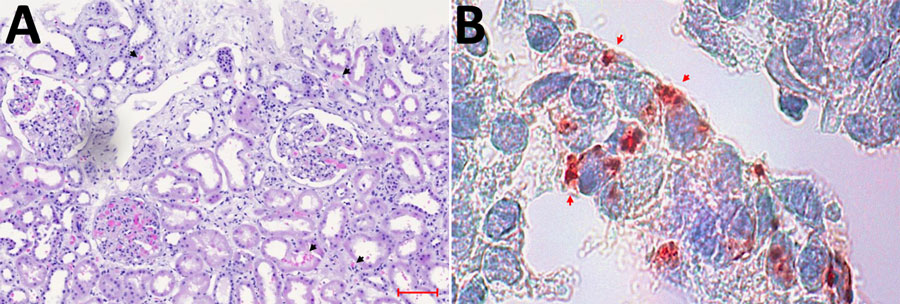
Figure 1. Light microscopic analysis and immunohistochemical staining results of index patient’s kidney biopsy indicating severe Seoul virus infection with acute kidney injury, Germany, March 2024. A) Light microscopy of the hematoxylin- and eosin-stained biopsy demonstrating severe acute tubular injury with interstitial and tubular bleeding (black arrow heads), tissue edema, and interstitial inflammation. Scale bar indicates 100 μm. B) Immunohistochemical staining (red) using mouse monoclonal antihantavirus antibody (Progen Biotechnik, https://www.progen.com) and the DCS-AEC chromogen kit (Agilent–DAKO, https://www.agilent.com) showing hantavirus antigen in renal tubular cells (red arrow heads). Original magnification ×100.
Main Article
Page created: September 02, 2025
Page updated: September 25, 2025
Page reviewed: September 25, 2025
The conclusions, findings, and opinions expressed by authors contributing to this journal do not necessarily reflect the official position of the U.S. Department of Health and Human Services, the Public Health Service, the Centers for Disease Control and Prevention, or the authors' affiliated institutions. Use of trade names is for identification only and does not imply endorsement by any of the groups named above.