Volume 31, Number 10—October 2025
Research Letter
Disseminated Blastomycosis Mimicking Tuberculosis, China
Can Guo, Yanjing Pan, Jiajia Yu, Linyan Yao, Yuhua He, Junwei Cui , Mengqiu Gao , and Yu Pang
Figure 1
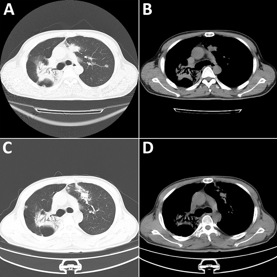
Figure 1. Chest computed tomography (CT) images from a patient with disseminated blastomycosis mimicking tuberculosis, China. A, B) Chest CT at first admission revealed bilateral lobar infiltrates (A) and patchy opacities (B). C, D) Chest CT taken 2 months later showed resolution of left lung lesions but progression of right lung lesions.
Page created: August 08, 2025
Page updated: September 25, 2025
Page reviewed: September 25, 2025
The conclusions, findings, and opinions expressed by authors contributing to this journal do not necessarily reflect the official position of the U.S. Department of Health and Human Services, the Public Health Service, the Centers for Disease Control and Prevention, or the authors' affiliated institutions. Use of trade names is for identification only and does not imply endorsement by any of the groups named above.