Skip directly to site content Skip directly to page options Skip directly to A-Z link Skip directly to A-Z link Skip directly to A-Z link

Volume 31, Number 12—December 2025

Dispatch

Pancreatic Schistosomiasis, China, 2020–2024

Long He1, Chuanbing Zhao1, Yu Lu1, Hongzhen Wei, Zanglong Deng, Yunpeng Zhang, and Tao YinComments to Author 
Author affiliation: Union Hospital Department of Pancreatic Surgery, Tongji Medical College, Huazhong University of Science and Technology, Wuhan, China

Main Article

Figure 1

Imaging from 4 cases of pancreatic schistosomiasis, China, 2020–2024. A, B) Computed tomography (CT) images from patient 1 revealing a mass involving the pancreas. C) Pathologic examination from fine-needle aspiration of patient 1 showing fibrinous exudate and schistosome eggs (arrows). D, E) Abdominal CT of patient 2 showing a nonenhancing, tubular lesion adjacent to the main pancreatic duct in the body of the pancreas. F) Pathologic examination of sample from patient 2 showing intraductal papillary mucinous neoplasm with moderate dysplasia and scattered schistosome eggs (arrows). G, H) Abdominal CT of patient 3 revealing a low-density mass in the pancreatic tail and multiple ring-enhancing hepatic nodules. I) Pathologic examination of sample from patient 3 showing schistosome egg deposition (arrows) with associated tissue necrosis.

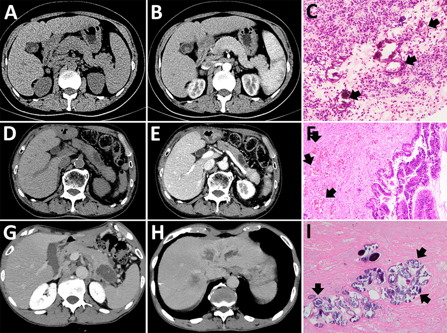
Figure 1. Imaging from 4 cases of pancreatic schistosomiasis, China, 2020–2024. A, B) Computed tomography (CT) images from patient 1 revealing a mass involving the pancreas. C) Pathologic examination from fine-needle aspiration of patient 1 showing fibrinous exudate and schistosome eggs (arrows). D, E) Abdominal CT of patient 2 showing a nonenhancing, tubular lesion adjacent to the main pancreatic duct in the body of the pancreas. F) Pathologic examination of sample from patient 2 showing intraductal papillary mucinous neoplasm with moderate dysplasia and scattered schistosome eggs (arrows). G, H) Abdominal CT of patient 3 revealing a low-density mass in the pancreatic tail and multiple ring-enhancing hepatic nodules. I) Pathologic examination of sample from patient 3 showing schistosome egg deposition (arrows) with associated tissue necrosis.

Main Article

1These authors are co–first authors.

Page created: November 18, 2025
Page updated: January 01, 2026
Page reviewed: January 01, 2026
The conclusions, findings, and opinions expressed by authors contributing to this journal do not necessarily reflect the official position of the U.S. Department of Health and Human Services, the Public Health Service, the Centers for Disease Control and Prevention, or the authors' affiliated institutions. Use of trade names is for identification only and does not imply endorsement by any of the groups named above.
file_external