Skip directly to site content Skip directly to page options Skip directly to A-Z link Skip directly to A-Z link Skip directly to A-Z link

Disclaimer: Early release articles are not considered as final versions. Any changes will be reflected in the online version in the month the article is officially released.

Volume 31, Number 3—March 2025
Research

Mycobacterium nebraskense Isolated from Patients in Connecticut and Oregon, USA

Mark L. MeterskyComments to Author , Ashley J. Losier, David A. Fraulino, Theodore A. Warnock, Cara D. Varley, Angela M. Le, Kevin L. Winthrop, John R. McArdle, Salika M. Shakir, and Reeti Khare
Author affiliation: University of Connecticut School of Medicine, Farmington, Connecticut, USA (M.L. Metersky, D.A. Fraulino, J.R. McArdle); Yale University School of Medicine, New Haven, Connecticut, USA (A.J. Losier); Oregon Health and Science University, Portland, Oregon, USA (T.A. Warnock, C.D. Varley, A.M. Le, K.L. Winthrop); Portland State University, Portland (T.A. Warnock, C.D. Varley, K.L. Winthrop); University of Utah ARUP Laboratories, Salt Lake City, Utah, USA (S.M. Shakir); National Jewish Health, Denver, Colorado, USA (R. Khare)

Main Article

Figure 1

High-resolution chest computed tomography scans from study of Mycobacterium nebraskense isolated from patients in Connecticut and Oregon, USA. A) Scan from case 2 in Connecticut showing bronchiectasis in the right middle and lower lobe, left lower lobe, and lingula and tree-in-bud nodularity in the lingula and right and left lower lobes of the lung. B) Scan slice from case 3 in Connecticut through the lower lung lobes showing 1 borderline dilated airway in the left lower lobe, performed in May 2019. C) Scan slice from case 3 at the same anatomic level showing numerous dilated airways and airway wall thickening in the left lower lobe of lung, performed in April 2020 soon after the first isolation of M. nebraskense. D) Scan from case 4 in Connecticut showing tree-in-bud nodularity in the right middle and lower lobes of the lung when patient had M. nebraskense isolated from her sputum in August 2020. E) Scan from case 4 in Connecticut showing resolution of the tree-in-bud nodularity in the right middle and lower lobes of the lung, performed in December 2022.

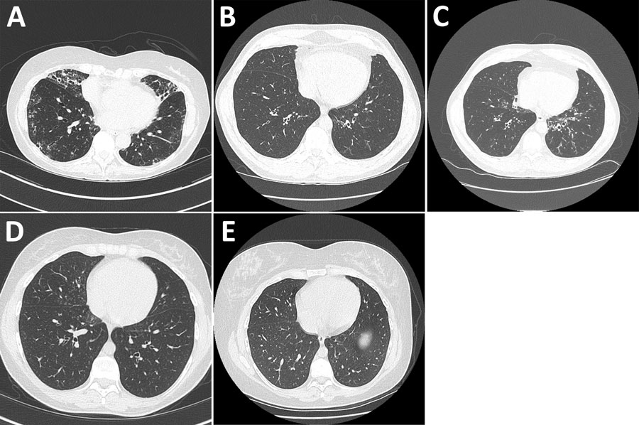
Figure 1. High-resolution chest computed tomography scans from study of Mycobacterium nebraskense isolated from patients in Connecticut and Oregon, USA. A) Scan from case 2 in Connecticut showing bronchiectasis in the right middle and lower lobe, left lower lobe, and lingula and tree-in-bud nodularity in the lingula and right and left lower lobes of the lung. B) Scan slice from case 3 in Connecticut through the lower lung lobes showing 1 borderline dilated airway in the left lower lobe, performed in May 2019. C) Scan slice from case 3 at the same anatomic level showing numerous dilated airways and airway wall thickening in the left lower lobe of lung, performed in April 2020 soon after the first isolation of M. nebraskense. D) Scan from case 4 in Connecticut showing tree-in-bud nodularity in the right middle and lower lobes of the lung when patient had M. nebraskense isolated from her sputum in August 2020. E) Scan from case 4 in Connecticut showing resolution of the tree-in-bud nodularity in the right middle and lower lobes of the lung, performed in December 2022.

Main Article

Page created: December 16, 2024
Page updated: February 21, 2025
Page reviewed: February 21, 2025
The conclusions, findings, and opinions expressed by authors contributing to this journal do not necessarily reflect the official position of the U.S. Department of Health and Human Services, the Public Health Service, the Centers for Disease Control and Prevention, or the authors' affiliated institutions. Use of trade names is for identification only and does not imply endorsement by any of the groups named above.
file_external