Volume 31, Number 5—May 2025
Dispatch
Tropheryma whipplei Infections, Mexico, 2019–2021
Jesús Delgado-de la Mora1 , Peter Grube-Pagola1, Christopher D. Paddock, Marlene DeLeon-Carnes, Alvaro C. Laga, Isaac H. Solomon, José María Remes-Troche, Jesús Javier Baquera-Heredia, Gabriel Quintero-Bustos, Juan Carlos León-Contreras, Arturo Ángeles-Ángeles, and Braulio Martínez-Benítez
Figure 1
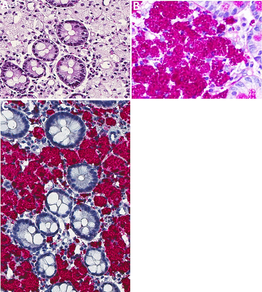
Figure 1. Microscopic and immunohistochemical examination of duodenal tissue samples from a 63-year-old man with Tropheryma whipplei infection, Mexico, 2019. A, B) Hematoxylin and eosin–stained tissue. Microscopic examination showed abundant macrophages in the lamina propria with foamy cytoplasm (A; original magnification ×10); and intracytoplasmic inclusions that stain with PAS (B; original magnification ×40). C) Immunohistochemistry reaction for T. whipplei showed intense positivity in the cytoplasmatic inclusions (original magnification ×40).
1These authors contributed equally to this article.
Page created: March 10, 2025
Page updated: April 25, 2025
Page reviewed: April 25, 2025
The conclusions, findings, and opinions expressed by authors contributing to this journal do not necessarily reflect the official position of the U.S. Department of Health and Human Services, the Public Health Service, the Centers for Disease Control and Prevention, or the authors' affiliated institutions. Use of trade names is for identification only and does not imply endorsement by any of the groups named above.