Volume 31, Number 6—June 2025
Etymologia
Eschar [esʹ kahr, esʹ kǝr]
Cite This Article
Citation for Media
Eschars, distinctive skin lesions from rickettsial infection induced by feeding ticks or mites, play a crucial role in diagnosing scrub typhus and spotted fever rickettsioses. Eschars occur at the sites where rickettsial pathogens are inoculated into the skin by an arthropod vector and typically emerge within a few (median 5) days after tick or mite bites, during the incubation period before symptom onset.
Figure
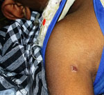
Figure. An eschar in the axillary area in the stage of healing in a patient with diagnosed scrub typhus.
Rickettsia-associated eschar is caused by rickettsial growth in endothelial cells, leading to thrombosis, ischemia, and necrosis of dermal tissue. The lesion initially resembles a small papule or pustule, then evolves into a central 0.5–3.0-cm ulcer covered by brown-black crust and encircled by a red halo (Figure), which can take several weeks to heal and can leave a small, depressed scar. Rickettsial eschars are typically painless, nonitchy, and frequently overlooked in patients with dark complexions, resulting in delayed diagnosis and treatment. PCR detection of pathogen DNA from eschar tissue or swab samples serves as a rapid diagnostic tool in early stages of eschar-associated rickettsial infection.
Sometimes, atypical eschars resembling acne may be observed. In mite-associated rickettsioses, eschars appear in areas where skin surfaces meet or clothes bind, such as the axilla, groin, neck, and waist, but occasionally occur at uncommon sites, such as wrist or elbow joints. However, tick-associated rickettsioses often manifest eschars on the extremities and trunk. Cutaneous manifestations of other infectious diseases, including tularemia, leishmaniasis, anthrax, and certain mycobacterial and fungal infections, can also produce eschars.
The term eschar finds its root from the Ancient Greek eskhára, meaning hearth, brazier, or scab, from which Middle French eschare and Late Latin eschara, both signifying scar or scab, evolved. That linguistic journey reflects the lesion’s historical association with healing and scarring of burnt skin.
Acknowledgment
We extend our sincere thanks to Rohit Beniwal and Asif Kavathekar for their support.
References
- Bechah Y, Socolovschi C, Raoult D. Identification of rickettsial infections by using cutaneous swab specimens and PCR. Emerg Infect Dis. 2011;17:83–6. DOIPubMedGoogle Scholar
- Jensenius M, Fournier PE, Kelly P, Myrvang B, Raoult D. African tick bite fever. Lancet Infect Dis. 2003;3:557–64. DOIPubMedGoogle Scholar
- Kim DM, Won KJ, Park CY, Yu KD, Kim HS, Yang TY, et al. Distribution of eschars on the body of scrub typhus patients: a prospective study. Am J Trop Med Hyg. 2007;76:806–9. DOIPubMedGoogle Scholar
- Merriam Webster.com Dictionary. Eschar [cited 2024 Feb 8]. https://www.merriam-webster.com/dictionary/eschar
- Paddock CD, Finley RW, Wright CS, Robinson HN, Schrodt BJ, Lane CC, et al. Rickettsia parkeri rickettsiosis and its clinical distinction from Rocky Mountain spotted fever. Clin Infect Dis. 2008;47:1188–96. DOIPubMedGoogle Scholar
Figure
Cite This ArticleOriginal Publication Date: May 20, 2025
Related Links
Table of Contents – Volume 31, Number 6—June 2025
| EID Search Options |
|---|
|
|
|
|
|
|
Please use the form below to submit correspondence to the authors or contact them at the following address:
Mahima Mittal, Department of Pediatrics, All India Institute of Medical Sciences, Kaysa Rd, Gorakhpur, Uttar Pradesh 273008, India
Top