Volume 31, Number 6—June 2025
Dispatch
Cadaveric Human Growth Hormone–Associated Creutzfeldt-Jakob Disease with Long Latency Period, United States
Anatevka S. Ribeiro , Andrew B. Wolf, Ellen W. Leschek, Lawrence B. Schonberger, Joseph Y. Abrams, Ryan A. Maddox, Brian S. Appleby, Katie Glisic, Aaron Carlson, and Elizabeth Matthews
Figure 1
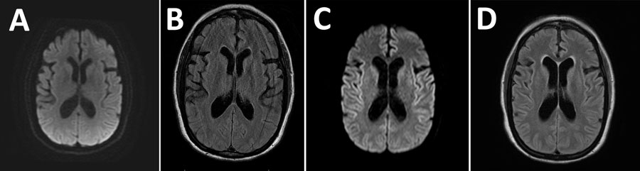
Figure 1. Magnetic resonance imaging of the brain from a case of cadaveric human growth hormone–associated Creutzfeldt-Jakob disease with long latency period, United States. A, B) Images obtained at initial clinical examination were unremarkable. C, D) Images obtained 3 months later demonstrated subtle areas of symmetric T2 hyperintensity in the insulae and frontotemporal lobes (C) and deep gray structures with diffusion restriction along the bilateral insulae and caudate heads without gadolinium enhancement (D).
Page created: April 16, 2025
Page updated: May 27, 2025
Page reviewed: May 27, 2025
The conclusions, findings, and opinions expressed by authors contributing to this journal do not necessarily reflect the official position of the U.S. Department of Health and Human Services, the Public Health Service, the Centers for Disease Control and Prevention, or the authors' affiliated institutions. Use of trade names is for identification only and does not imply endorsement by any of the groups named above.