Volume 31, Number 6—June 2025
Research Letter
Baylisascaris procyonis Roundworm in Common Raccoon (Procyon lotor), Mexico
Figure 1
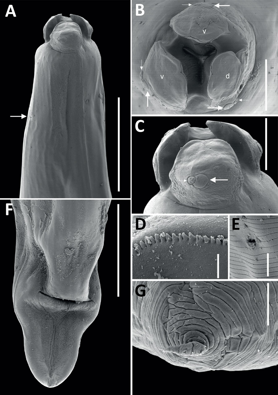
Figure 1. Scanning electron micrographs of female Baylisascaris procyonis roundworm detected in common raccoon (Procyon lotor), Mexico. A) Anterior end of worm body. Arrow indicates the excretory pore. Scale bar indicates 400 μm. B) Cephalic end, apical view of worm. Lowercase letters indicate dorsal (d) and ventral (v) lips, each having 1 large double papillae instead of a dorsal lip with 2 large double papillae, as reported previously (4). Small and large arrows indicate the double papillae. Scale bar indicates 100 μm. C) Cephalic end of worm. Small and large arrows indicate the double papillae. Scale bar indicates 100 μm. D) Magnified image of labial denticles. Scale bar indicates 5 μm. E) Magnified image of the excretory pore. Scale bar indicates 10 μm. F) Posterior end of the worm body. Scale bar indicates 300 μm. G) Magnified image of the tail tip. Scale bar indicates 10 μm.
References
- US Geological Survey, National Wildlife Health Center. Baylisascaris larva migrans. Circular 1412. 2016 [cited 2024 Apr 25]. https://pubs.usgs.gov/circ/1412/cir1412.pdf
- Bauer C. Baylisascariosis—infections of animals and humans with ‘unusual’ roundworms. Vet Parasitol. 2013;193:404–12. DOIPubMedGoogle Scholar
- Lipton BA, Oltean HN, Capron RB, Hamlet A, Montgomery SP, Chancey RJ, et al. Baylisascaris procyonis roundworm infection in child with autism spectrum disorder, Washington, USA, 2022. Emerg Infect Dis. 2023;29:1232–5. DOIPubMedGoogle Scholar
- Gu XH, Chen HX, Hu JJ, Li L. Morphology and ASAP analysis of the important zoonotic nematode parasite Baylisascaris procyonis (Stefahski and Zarnowski, 1951), with molecular phylogenetic relationships of Baylisascaris species (Nematoda: Ascaridida). Parasitology. 2024;151:200–12.PubMedGoogle Scholar
- Pinacho-Pinacho CD, Calixto-Rojas M, García-Vásquez A, Guzmán-Valdivieso I, Barrios-Gutiérrez JJ, Rubio-Godoy M. Species delimitation of Gyrodactylus (Monogenea: Gyrodactylidae) infecting the southernmost cyprinids (Actinopterygii: Cyprinidae) in the New World. Parasitol Res. 2021;120:831–48. DOIPubMedGoogle Scholar
- García-Varela M, Nadler SA. Phylogenetic relationships of Palaeacanthocephala (Acanthocephala) inferred from SSU and LSU rDNA gene sequences. J Parasitol. 2005;91:1401–9. DOIPubMedGoogle Scholar
- Tamura K, Stecher G, Kumar S. MEGA11: molecular evolutionary genetics analysis version 11. Mol Biol Evol. 2021;38:3022–7. DOIPubMedGoogle Scholar
- Baldi M, Alvarado G, Smith S, Santoro M, Bolaños N, Jiménez C, et al. Baylisascaris procyonis parasites in raccoons, Costa Rica, 2014. Emerg Infect Dis. 2016;22:1502–3. DOIPubMedGoogle Scholar
- Mezhua-Velázquez MJ, Serna-Lagunes R, Torres-Cantú GB, Pérez-Gracida LD, Salazar-Ortiz J, Mora-Collado N. Diversity of medium and large mammals from the Ejido Zomajapa Zongolica, Veracruz, Mexico: management implications [in Spanish.]. Ecosist Recur Agropecu. 2022;9:
e3316 . - García-Padilla BF, Zalapa SS, Guerrero-Vázquez S, Pérez-Arteaga A, Camacho-Rodríguez A. Abundance and activity of domestic and wild medium-sized mammals in a protected mangrove remnant, Puerto Vallarta, Mexico. West N Am Nat. 2021;81:285–92. DOIGoogle Scholar