Volume 31, Number 7—July 2025
Research Letter
Syphilitic Aortitis with Concomitant Neurosyphilis in Asymptomatic Patient
Evan Czulada , Quinn Seau, Tyler Geshay, Danny Rayes, Trevor Wyand, Ethan Fraser, Ronald Beaulieu, and Justin Beckett
Figure
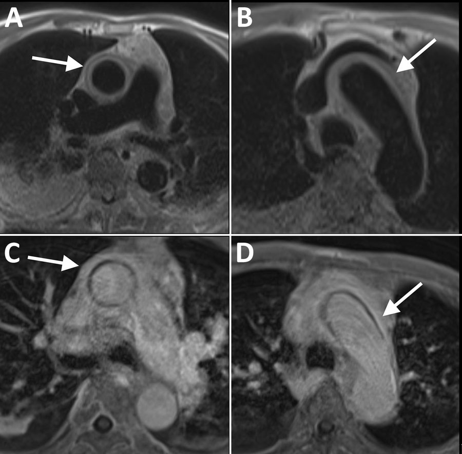
Figure. Electrocardiogram-gated magnetic resonance imaging of the ascending aorta in an 89-year-old patient with syphilitic aortitis and concomitant neurosyphilis, Washington, DC, USA. A, B) A circumferential periaortic T2 hyperintense signal was depicted at the main pulmonary artery (A) and aortic arch (B) on black blood prepared half-Fourier acquisition single-shot turbo spin-echo sequence images (white arrows). C, D) Contrast-enhanced T1-weighted magnetic resonance images at the main pulmonary artery (C) and aortic arch (D) show wall thickening and enhancement (white arrows) compatible with aortitis.
Page created: June 06, 2025
Page updated: June 25, 2025
Page reviewed: June 25, 2025
The conclusions, findings, and opinions expressed by authors contributing to this journal do not necessarily reflect the official position of the U.S. Department of Health and Human Services, the Public Health Service, the Centers for Disease Control and Prevention, or the authors' affiliated institutions. Use of trade names is for identification only and does not imply endorsement by any of the groups named above.