Skip directly to site content Skip directly to page options Skip directly to A-Z link Skip directly to A-Z link Skip directly to A-Z link

Disclaimer: Early release articles are not considered as final versions. Any changes will be reflected in the online version in the month the article is officially released.

Volume 31, Number 8—August 2025

Dispatch

Microsporidial Keratoconjunctivitis Caused by Vittaforma corneae, Sea of Galilee, Israel, 2022–2024

Asaf Friehmann, Irit Lubitz, Fidaa El Zhalka, and Sharon AmitComments to Author 
Author affiliation: Meir Medical Center, Kfar Sava, Israel (A. Friehmann, F. El Zhalka); Tel-Aviv University, Tel-Aviv, Israel (A. Friehmann, S. Amit); Sheba Medical Center, Ramat-Gan, Israel (I. Lubitz, S. Amit).

Main Article

Figure

Slit-lamp photos of 3 patients before and after treatment of microsporidial keratoconjunctivitis caused by Vittaforma corneae, Sea of Galilee, Israel, 2022–2024. A) Patient 1 before treatment, visual acuity 0.8 decimal. B) Patient 2 before treatment, visual acuity 0.3 decimal. C) Patient 3 before treatment, visual acuity 0.3 decimal. D) Patient 1 after treatment, visual acuity 1 decimal. E) Patient 2 after treatment, visual acuity 1 decimal. F) Patient 3 after treatment, visual acuity 1 decimal. Clinical manifestations in all 3 patients included corneal epithelial microsporidial infiltrates and conjunctival irritation. After treatment with 0.02% topical chlorhexidine, the infiltrates resolved without scarring or other complications.

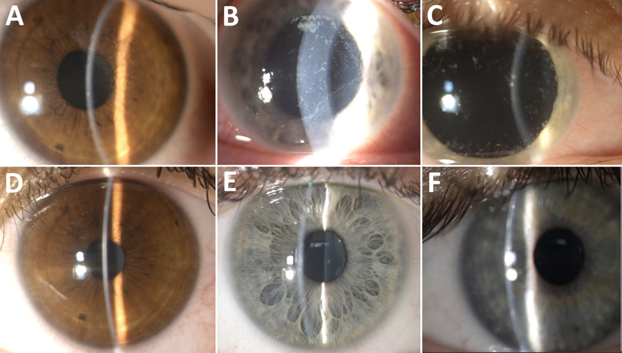
Figure. Slit-lamp photos of 3 patients before and after treatment of microsporidial keratoconjunctivitis caused by Vittaforma corneae, Sea of Galilee, Israel, 2022–2024. A) Patient 1 before treatment, visual acuity 0.8 decimal. B) Patient 2 before treatment, visual acuity 0.3 decimal. C) Patient 3 before treatment, visual acuity 0.3 decimal. D) Patient 1 after treatment, visual acuity 1 decimal. E) Patient 2 after treatment, visual acuity 1 decimal. F) Patient 3 after treatment, visual acuity 1 decimal. Clinical manifestations in all 3 patients included corneal epithelial microsporidial infiltrates and conjunctival irritation. After treatment with 0.02% topical chlorhexidine, the infiltrates resolved without scarring or other complications.

Main Article

Page created: June 13, 2025
Page updated: July 02, 2025
Page reviewed: July 02, 2025
The conclusions, findings, and opinions expressed by authors contributing to this journal do not necessarily reflect the official position of the U.S. Department of Health and Human Services, the Public Health Service, the Centers for Disease Control and Prevention, or the authors' affiliated institutions. Use of trade names is for identification only and does not imply endorsement by any of the groups named above.
file_external