Volume 31, Number 9—September 2025
Dispatch
Detection of Rat Lungworm (Angiostrongylus cantonensis) in Rats and Gastropods, Italy
Cite This Article
Citation for Media
Abstract
The emerging zoonotic nematode Angiostrongylus cantonensis causes severe neural angiostrongyliasis in both humans and animals. The parasite has been reported in Spain. We detected A. cantonensis in rats and gastropods from the Campania region, southern Italy, demonstrating its broad distribution on the southern coast of Europe.
The rat lungworm, Angiostrongylus cantonensis, a neurotropic zoonotic parasite, is receiving increasing attention because of its potential to cause severe neurologic disease in humans and animals (1). This rat lungworm has an indirect life cycle involving rats (mainly Rattus spp.) as definitive hosts, mollusks as intermediate hosts, and different paratenic and transient hosts such as frogs, lizards, and crustaceans (2). Infection in humans usually occurs by accidental ingestion of infective third-stage larvae (L3) found in raw or undercooked snails or paratenic hosts or by contact with L3-contaminated water or products (3). Identified in China in 1935, A. cantonensis has since become endemic in Southeast Asia, East Asia, North and South America, and selected Pacific and Caribbean islands, where most human cases of neuroangiostrongyliasis occur; >7,000 human cases have been recorded worldwide (4).
In the past 2 decades, the geographic range of A. cantonensis lungworms has increased in Europe, and they have been detected in the Canary Islands (Tenerife, Spain) (5), in the Balearic Islands (Mallorca, Spain) (6), and most recently in mainland Spain (Valencia) (7), indicating a continued spread in the Mediterranean basin. Although human cases remain rare in Europe and have been associated with travel to well-established endemic regions, such as Southeast Asia and the Caribbean Islands (8), the subtropical climate and historically active maritime trade in Naples, Italy, provide favorable conditions for the spread of A. cantonensis to human and animal hosts. We investigated Rattus spp. rats and snail populations in periurban and rural areas of the Campania region of southern Italy to determine whether the A. cantonensis lungworm has spread to this region along the Mediterranean Coast of Europe.
Figure 1

Figure 1. Sampling locations used for detection of rat lungworm (Angiostrongylus cantonensis) in rats and gastropods, Italy. Solid black circles indicate sites from which A. cantonensis–positive samples were...
We obtained a total of 32 frozen rat specimens, 10 R. rattus and 22 R. norvegicus, from a pest control company operating in metropolitan Naples and its surroundings. We conducted an initial sampling phase randomly across various locations. After we detected A. cantonensis lungworm in rats, we conducted a second sampling, during which we collected 352 gastropods from locations where infected rats were collected and from nearby areas selected at random. In total, we sampled rodent and gastropod samples from 15 locations during January–November 2024 (Figure 1; Appendix 1).
We necropsied rats at the Experimental Zooprophylactic Institute of Southern Italy in Naples. We isolated the heart and lungs and inspected them for adult Angiostrongylus spp. worms, characterized by the distinctive barber pole appearance in female nematodes; we preserved isolated worms in 96% ethanol. We froze tissue samples from the brain, heart, kidneys, liver, lungs, and spleen of rats for molecular analysis. We confirmed rat species on the basis of DNA extracted from spleen samples by using the Nucleic Acid Extraction Kit (Magnetic Bead Method) (Zybio, https://www.zybio.com), followed by amplification of the mitochondrial cytochrome b gene (Appendix 2). We detected A. cantonensis nematodes in 13 (40.6%) of 32 rats collected from 3 locations, with a mean of 7 (range 1–24) worms per rat (Table; Figure 1; Appendix 1).
Figure 2
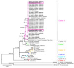
Figure 2. Maximum-likelihood phylogenetic tree of rat lungworm (Angiostrongylus cantonensis) detected in rats and gastropods, Italy. Tree is based on cytochrome c oxidase subunit 1 gene (1,578 bp) and partial...
Molecular analysis confirmed A. cantonensis nematodes in 12 positive rats, and we subsequently sequenced 69 adult worms. We extracted DNA from those adult worms by using the DNeasy Blood & Tissue Kit (QIAGEN, https://www.qiagen.com), and we amplified the complete cytochrome c oxidase subunit 1 (CO1) gene (Appendix 2). The obtained sequences revealed 2 distinct haplotypes (NAP1 and NAP2), differing by 2 single-nucleotide polymorphisms (SNPs) at positions 1092 and 1481 of the CO1 gene; the SNP at position 1481 resulted in a different amino acid. In a maximum-likelihood phylogenetic tree (Figure 2), both haplotypes clustered within clade II sensu, as previously defined (9), alongside other sequences from Europe, except for 1 (GenBank accession no. PP468354; 215 bp) from Valencia that clustered in a separate clade, a sister to clade I, differing from all other sequences from Europe in 3 SNPs. Compared with the TEN.1 isolate (GenBank accession no. MK570629) from Tenerife, Spain, each haplotype from Italy differed by a single SNP: NAP1, detected in Naples (Porto) and Marano di Napoli, differed at position 1092; and NAP2, detected in Naples (Vomero), differed at position 1481. The 394-bp sequence from Mallorca, Spain (GenBank accession no. MN227185), was identical to NAP2. Among the Valencia isolates, 10 sequences were identical to NAP1, 3 were identical to NAP2, and the rest differed in 1, 2, or 3 SNPs from the other sequences from Italy.
Collected gastropods were identified to species level by a trained malacologist on the basis of morphological criteria. Molecular identification was performed on juvenile and shell-less specimens lacking distinct morphological characteristics. DNA was extracted from muscle tissue using the same protocol used for rat spleen samples, with an extended overnight prelysis phase at 56°C, optimized for the L3 stage of A. cantonensis. Molecular identification was made on the basis of sequences of the mitochondrial 16S rRNA gene (Appendix 2). We detected A. cantonensis worms by using a species-specific quantitative PCR on DNA isolated from gastropod tissue (11). Of the 352 gastropods examined, 8 (2.3%) gastropods from 2 localities tested positive for A. cantonensis DNA (Figure 1, Table; Appendix 2). We successfully obtained 6 CO1 gene fragment sequences from the 8 positive gastropods (Appendix 2), and compared those with sequences from adult A. cantonensis lungworms from rats in this study. All sequences belonged to the NAP1 haplotype.
We provide robust evidence that the A. cantonensis rat lungworm is in the central Mediterranean region in the Naples area of Italy. Circulation of this zoonotic nematode in the highly populated Naples metropolitan area is concerning because of its ability to cause severe neurologic and ocular disorders in humans. Because Naples has an environment ideal for A. cantonensis transmission to the human population, enhanced awareness is needed among healthcare practitioners and diagnostic protocols should be revised and applied locally in the differential diagnosis of meningoencephalitis cases (12). In addition, considering reported clinical cases in domestic animals and in wildlife in known endemic foci (13–15), veterinary practitioners in the Naples area should be alerted.
Mr. Pandian is a doctoral student at the Department of Veterinary Sciences of Czech University of Life Sciences Prague. His research interests include all aspects of emergence and circulation of Angiostrongylus cantonensis in endemic and newly formed foci.
Acknowledgments
We thank Petr Janoš for preparing the map of localities studied. We also thank Radovan Coufal and Veronika Horsáková for their assistance with the morphologic and molecular identification of gastropod specimens.
This study was supported by the Czech Science Foundation, grant no. 22-26136S. The work of Divakaran Pandian was supported by the Erasmus+ mobility program, and Anna Šipková was supported by Specific—Support of Student Projects (no. MUNI/A/1762/2024).
References
- Morgan ER, Modry D, Paredes-Esquivel C, Foronda P, Traversa D. Angiostrongylosis in animals and humans in Europe. Pathogens. 2021;10:1236. DOIPubMedGoogle Scholar
- Alicata JE. Biology and distribution of the rat lungworm, Angiostrongylus cantonensis, and its relationship to eosinophilic meningoencephalitis and other neurological disorders of man and animals. Adv Parasitol. 1965;3:223–48. DOIPubMedGoogle Scholar
- Cowie RH. Biology, systematics, life cycle, and distribution of Angiostrongylus cantonensis, the cause of rat lungworm disease. Hawaii J Med Public Health. 2013;72(Suppl 2):6–9.PubMedGoogle Scholar
- Turck HC, Fox MT, Cowie RH. Paratenic hosts of Angiostrongylus cantonensis and their relation to human neuroangiostrongyliasis globally. One Health. 2022;15:
100426 . DOIPubMedGoogle Scholar - Foronda P, López-González M, Miquel J, Torres J, Segovia M, Abreu-Acosta N, et al. Finding of Parastrongylus cantonensis (Chen, 1935) in Rattus rattus in Tenerife, Canary Islands (Spain). Acta Trop. 2010;114:123–7. DOIPubMedGoogle Scholar
- Paredes-Esquivel C, Sola J, Delgado-Serra S, Puig Riera M, Negre N, Miranda MÁ, et al. Angiostrongylus cantonensis in North African hedgehogs as vertebrate hosts, Mallorca, Spain, October 2018. Euro Surveill. 2019;24:33. DOIPubMedGoogle Scholar
- Galán-Puchades MT, Gómez-Samblás M, Osuna A, Sáez-Durán S, Bueno-Marí R, Fuentes MV. Update on the first finding of the rat lungworm, Angiostrongylus cantonensis, in Rattus spp. in continental Europe, Valencia, Spain, 2022. Pathogens. 2023;12:4. DOIPubMedGoogle Scholar
- Federspiel F, Skovmand S, Skarphedinsson S. Eosinophilic meningitis due to Angiostrongylus cantonensis in Europe. Int J Infect Dis. 2020;93:28–39. DOIPubMedGoogle Scholar
- Trifinopoulos J, Nguyen LT, von Haeseler A, Minh BQ. W-IQ-TREE: a fast online phylogenetic tool for maximum likelihood analysis. Nucleic Acids Res. 2016;44(W1):
W232-5 . DOIPubMedGoogle Scholar - Tian X, Chen S, Duan L, Qian Y, Li H, Lv S. The global spread pattern of rat lungworm based on mitochondrial genetics. Pathogens. 2023;12:13. DOIPubMedGoogle Scholar
- Anettová L, Baláž V, Coufal R, Horsák M, Izquierdo-Rodriguez E, Šipková A, et al. Lizards as sentinels for the distribution of Angiostrongylus cantonensis. Epidemiol Infect. 2024;152:
e168 . DOIPubMedGoogle Scholar - Ansdell V, Kramer KJ, McMillan JK, Gosnell WL, Murphy GS, Meyer BC, et al. Guidelines for the diagnosis and treatment of neuroangiostrongyliasis: updated recommendations. Parasitology. 2021;148:227–33. DOIPubMedGoogle Scholar
- Garijo-Toledo M, Alarcón-Elbal PM, Montero E, Bravo-Barriga D, Sansano-Maestre J, Ahuir-Baraja AE, et al. Mortality associated with Angiostrongylus cantonensis in non-human primates in Europe. Int J Parasitol. 2025;55:427–34. DOIPubMedGoogle Scholar
- Odani J, Sox E, Coleman W, Jha R, Malik R. First documented cases of canine neuroangiostrongyliasis due to Angiostrongylus cantonensis in Hawaii. J Am Anim Hosp Assoc. 2021;57:42–6. DOIPubMedGoogle Scholar
- Cowie RH, Malik R, Morgan ER. Comparative biology of parasitic nematodes in the genus Angiostrongylus and related genera. Adv Parasitol. 2023;121:65–197. DOIPubMedGoogle Scholar
Figures
Table
Cite This ArticleOriginal Publication Date: August 19, 2025
1These first authors contributed equally to this article.
Table of Contents – Volume 31, Number 9—September 2025
| EID Search Options |
|---|
|
|
|
|
|
|
Please use the form below to submit correspondence to the authors or contact them at the following address:
David Modrý, Department of Veterinary Sciences, Faculty of Agrobiology, Food and Natural Resources, Czech University of Life Sciences, Kamýcká 129, 16500 Prague, Czech Republic
Top