Volume 32, Number 2—February 2026
Research Letter
Severe Respiratory Diphtheria-Like Illness Caused by Toxigenic Corynebacterium ulcerans
Cite This Article
Citation for Media
Abstract
We report a possible zoonotic case of severe diphtheria-like respiratory illness in Norway caused by a previously unreported toxigenic Corynebacterium ulcerans sequence type. This case highlights C. ulcerans as an emerging pathogen that can cause life-threatening disease. Clinicians should be aware of C. ulcerans infection, even in regions where diphtheria is rare.
Corynebacterium ulcerans bacteria can cause respiratory and nonrespiratory infections in humans, and respiratory diphtheria-like illness caused by toxigenic C. ulcerans is increasing (1). Globally, C. ulcerans bacteremia is rare, and limited clinical data are available. We describe a case of severe respiratory diphtheria-like illness caused by toxigenic C. ulcerans bacteremia in an immunocompromised patient in Norway without travel history.
On January 11, 2024, a 74-year-old immunocompromised man was referred to the emergency department for a 2-day history of breathlessness, productive cough, malaise, and cognitive impairment. He had a sore throat and cold-like symptoms 8–9 days earlier. He lived in an urban area with his wife and dog. Medical history included coronary disease, type 2 diabetes mellitus, chronic obstructive pulmonary disease with obstructive sleep apnea, and a hemicolectomy for cancer in 2019. He had been immunocompromised since 2021 from weekly methotrexate (20 mg) and etanercept (50 mg) for psoriatic arthritis.
At admission, he was afebrile but in respiratory distress with marked hoarseness and wheezing on auscultation; throat examination was unremarkable. He was hemodynamically stable; oxygen saturation was 88% on room air. Electrocardiograph showed sinus rhythm; echocardiography revealed preserved biventricular systolic function. Chest radiograph showed a retrocardiac infiltrate; laboratory findings indicated infection (Table).
He was treated with ampicillin (1 g 4×/d), prednisolone (40 mg), inhaled bronchodilators for suspected exacerbation of his pulmonary disease, and 2 L/min of oxygen via nasal cannula. Blood cultures taken at admission showed gram-positive rods after 27 hours using the BD Bactec FX system (Becton Dickinson, https://www.bd.com). MALDI BioTyper (Bruker Daltonik GmbH, https://www.bruker.com) identified C. ulcerans, prompting a diphtheria-like illness diagnosis. The Norwegian Institute of Public Health confirmed C. ulcerans using API Coryne version 4.0 system (bioMérieux, https://www.biomerieux.com) and by PCR. We also identified the tox gene by PCR and toxin production by a modified Elek test (2). We sequenced the isolate on a NextSeq platform (Illumina, https://www.illumina.com), and curators at BIGSdb-Pasteur MLST database (https://bigsdb.pasteur.fr) identified it as sequence type 1032.
Two days after admission, antibiotic therapy was changed to intravenous penicillin G ( 3 g 4×/d), and the patient received 100,000 units of diphtheria antitoxin. Respiratory distress worsened, and the patient was transferred to the intensive care unit. Four days after admission, ventricular tachycardia developed but was successfully cardioverted after intubation.
Figure
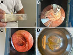
Figure. Clinical findings from a case of severe respiratory diphtheria-like illness caused by toxigenic Corynebacterium ulcerans, Norway. A) Pseudomembranes coughed up by the patient on day 1 of hospitalization....
The patient required mechanical ventilation for 7 days. Bronchoscopy revealed pseudomembranes in the lower airways, which were removed (Figure). He completed 14 days of antibiotic therapy, mainly benzylpenicillin, and 7 days of cefotaxime for ventilator-associated Staphylococcus aureus pneumonia.
An initial throat swab sample from hospitalization day 2 was C. ulcerans–negative, but follow-up samples from days 5 and 8 were C. ulcerans–positive. Follow-up samples on days 18 and 19 were negative, and the patient’s isolation was discontinued; he was discharged after 23 days. Comparison of antibody levels against diphtheria toxoid showed a protective level in 2020 (0.18 IU/mL) and >3.0 IU/mL on hospitalization day 7.
At 2-month follow-up, the patient had bilateral thigh numbness. Electroneurography confirmed sensorimotor polyneuropathy.
Although severe C. ulcerans infections have been described (3,4), detailed clinical data on patients with positive blood cultures remain rare. Bacteremia appears linked to severe immunosuppression (3,5). This patient had protective diphtheria antibody titers but fulminant illness developed, requiring ventilation, antibiotics, and diphtheria antitoxin. A similar pattern was observed during a diphtheria epidemic in Russia in the 1990s (6). Although nontoxigenic C. diphtheriae can cause severe disease, diphtheria toxin remains the main virulence factor, inhibiting protein synthesis and causing cell death, especially in myocardium and peripheral nerves (7). This patient’s ventricular arrhythmia and subsequent polyneuropathy were consistent with diphtheria toxin–mediated toxicity (7).
Toxigenic C. ulcerans transmission remains uncertain. Although primarily zoonotic, with dairy cattle as a classic reservoir (1), infections from domestic and wild animals have been reported (3,4). We suspected but could not confirm transmission from this patient’s dog because no microbial samples were collected. The dog showed no signs of respiratory or skin disease before the patient’s hospitalization and remained in good health.
Because C. ulcerans among animals is not notifiable in Norway, no official data or surveillance results are available. The sequence type 1032 variant had not been previously reported in humans and has since been identified in 1 other sample from another patient in Norway 6 months later; however, the 2 patients had no known epidemiologic link (V. Skogen, unpub. data). Whole-genome sequence analysis revealed substantial genomic differences between the 2 patients’ isolates, supporting separate infection sources. Because potential human-to-human transmission has been suggested (8–10), we applied isolation precautions, but no secondary cases were detected among close contacts or healthcare workers.
In summary, diagnosing diphtheria-like illness in low-prevalence settings is difficult because selective media are rarely used, and standard respiratory culture media might not detect Corynebacterium spp. bacteria. In this case, positive blood culture aided in early recognition, an uncommon but critical finding because classic diphtheria symptoms developed. This case underscores that C. ulcerans is an emerging zoonotic pathogen capable of causing life-threatening disease and highlights that early microbiological diagnosis is crucial, even in regions where diphtheria is rare.
Dr. Helleren is a specialist in internal medicine and infectious diseases and is the head of the infectious diseases section at Sørlandet Hospital Kristiansand, Norway. Her current research interest is outpatient parenteral antimicrobial therapy.
References
- Hacker E, Antunes CA, Mattos-Guaraldi AL, Burkovski A, Tauch A. Corynebacterium ulcerans, an emerging human pathogen. Future Microbiol. 2016;11:1191–208. DOIPubMedGoogle Scholar
- World Health Organization. WHO laboratory manual for the diagnosis of diphtheria and other related infections. Geneva: The Organization; 2021.
- Yamamoto A, Hifumi T, Ato M, Iwaki M, Senoh M, Hatanaka A, et al. Clinical characteristics of Corynebacterium ulcerans infection, Japan. Emerg Infect Dis. 2023;29:1505–15. DOIPubMedGoogle Scholar
- Lartigue MF, Monnet X, Le Flèche A, Grimont PA, Benet JJ, Durrbach A, et al. Corynebacterium ulcerans in an immunocompromised patient with diphtheria and her dog. J Clin Microbiol. 2005;43:999–1001. DOIPubMedGoogle Scholar
- Bonmarin I, Guiso N, Le Flèche-Matéos A, Patey O, Patrick AD, Levy-Bruhl D. Diphtheria: a zoonotic disease in France? Vaccine. 2009;27:4196–200. DOIPubMedGoogle Scholar
- Danilova E, Jenum PA, Skogen V, Pilnikov VF, Sjursen H. Antidiphtheria antibody responses in patients and carriers of Corynebacterium diphtheriae in the Arkhangelsk region of Russia. Clin Vaccine Immunol. 2006;13:627–32. DOIPubMedGoogle Scholar
- Sharma NC, Efstratiou A, Mokrousov I, Mutreja A, Das B, Ramamurthy T. Diphtheria. Nat Rev Dis Primers. 2019;5:81. DOIPubMedGoogle Scholar
- Wagner KS, White JM, Crowcroft NS, De Martin S, Mann G, Efstratiou A. Diphtheria in the United Kingdom, 1986-2008: the increasing role of Corynebacterium ulcerans. Epidemiol Infect. 2010;138:1519–30. DOIPubMedGoogle Scholar
- Othieno R, Mark K, Etherson M, Foster G, Murray S, Kalima P, et al. A case of cutaneous toxigenic Corynebacterium ulcerans likely acquired from a domestic dog. Access Microbiol. 2019;1:
e000025 . DOIPubMedGoogle Scholar - Metz AR, White A, Ripplinger J, Spence Davizon E, Barnes M, Bauer M, et al. Notes from the field: toxigenic Corynebacterium ulcerans in humans and household pets—Utah and Colorado, 2022–2023. MMWR Morb Mortal Wkly Rep. 2024;73:534–5. DOIPubMedGoogle Scholar
Figure
Table
Cite This ArticleOriginal Publication Date: February 09, 2026
Table of Contents – Volume 32, Number 2—February 2026
| EID Search Options |
|---|
|
|
|
|
|
|
Please use the form below to submit correspondence to the authors or contact them at the following address:
Vegard Skogen, Faculty of Health Sciences, University of Tromsø, The Arctic University of Norway, PO Box 6050 Langnes, N-9037 Tromsø, Norway
Top