Volume 32, Number 9—September 2026
Dispatch
Global Spread of Trichophyton indotineae Tracked by Mass Spectrometry Identification Network, 2022–2025
 , and MIND Study Group,1
Cite This Article
Citation for Media
Abstract
Trichophyton indotineae is an emerging dermatophyte that is often resistant to terbinafine. We analyzed data produced by the Mass Spectrometry Identification global network. Those data revealed a 3-fold increase in T. indotineae identification during 2022–2025, demonstrating that this application is an effective tool for monitoring global spread of this pathogen.
Infections caused by the dermatophyte Trichophyton indotineae have recently emerged as a global public health concern (1). Although the first cases of infection by T. indotineae were retrospectively identified as early as 2008, beginning in 2016 dermatologists in India began to report a sharp increase in cases of extensive and difficult-to-treat dermatophytosis in their country. Those cases were later attributed to strains belonging to the T. mentagrophytes complex, which were frequently resistant to terbinafine (1). In 2020, the designation T. indotineae was proposed to denote these strains, although debate is ongoing about whether this clade should be considered a distinct species or merely a variety within T. mentagrophytes (2). Current alternative denominations include T. mentagrophytes internal transcribed spacer (ITS) genotype VIII and T. mentagrophytes variant. indotineae. T. indotineae infections are now being reported in many countries, mostly as individual case reports or small single-center series, illustrating the rapid international spread of this species through migration and travel. Only a few publications provide data at broader (regional or national) scales and over longer periods (3–5).
Macro-microscopic–based identification of T. indotineae remains challenging because of its close similarity with T. interdigitale and T. mentagrophytes. Reference identification relies on sequencing of the ITS1–5.8s–ITS2 region. Matrix-assisted laser desorption/ionization time-of-flight mass spectrometry has become established in recent years as a highly effective method for identifying fungal pathogens. The Mass Spectrometry Identification (MSI) application (https://msi.happy-dev.fr) is a free tool developed and hosted by the Parasitology-Mycology Laboratory of Hôpital Universitaire Pitié Salpêtrière (Paris, France) that enables accurate and fast identification of large numbers of fungal pathogens on the basis of their mass spectra (6). It includes a large number of reference spectra (17,563 spectra corresponding to 1,700 fungal species) and enables the identification of rare species. In March 2022, MSI was upgraded to identify T. indotineae with excellent sensitivity and specificity (>95% for both parameters) (7,8). The ability to produce a reliable identification relies on the presence of 2 pairs of peaks, the presence or absence of which enables T. interdigitale and T. mentagrophytes to be effectively distinguished (8). Furthermore, the widespread use of this free online application has enabled the creation of an extensive international network of laboratories with ≈450 regular users in 27 countries. In this study, we analyzed identification data generated by MSI to describe the spatial and temporal dynamics of T. indotineae infections.
We analyzed all spectra identified as T. indotineae during March 2022–December 2025 by MSI users. We considered only first-intention identifications with a score of >20 (i.e., above the threshold used for the application for correct species identification as previously described [9]). When the same spectrum was uploaded multiple times, only 1 entry was retained. We addressed the bias caused by the increase in the number of users over time by normalizing the number of T. indotineae identifications relative to the number of identifications for the genus Trichophyton spp. and for the T. mentagrophytes complex (i.e., T. mentagrophytes, T. interdigitale, and T. indotineae).
Figure 1
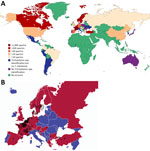
Figure 1. Geographic distribution of Trichophyton indotineae spectra in study of global spread of T. indotineaetracked by the Mass Spectrometry Identification (MSI) Network, 2022–2025. Maps indicate global (A)...
During the study period, 566 users from 54 countries submitted 1,657,334 spectra that led to the identification of fungal agents. Those identifications included 123,025 (7.7%) that were identified as Trichophyton spp. (by 316 users in 44 countries) and 3,399 (0.2%) that were identified as T. indotineae (by 183 users in 29 countries) (Figure 1, panel A). The number of T. indotineae identifications per user ranged from 1 to 299 spectra. Europe accounted for 63.8% of users, ahead of Asia (16.8%), Latin America (15.7%), North America (1.6%), Oceania (1.6%), and Africa (0.5%). As a consequence, most T. indotineae spectra were identified in Europe (3,040 [89.4%]). In western and northern Europe, which had the highest number of MSI users, detections were not limited to major urban centers.
Figure 2
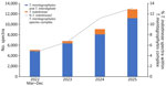
Figure 2. Temporal evolution of Trichophyton indotineae spectra identified in study of global spread of T. indotineaetracked by the Mass Spectrometry Identification Network, 2022–2025. Spectra were identified during...
Over the study period, the number of T. indotineae spectra increased continuously: from 237 in 2022 to 454 in 2023, then to 1,016 in 2024, and finally to 1,692 in 2025. At the same time, the percentage of spectra identified as T. indotineae within the Trichophyton genus increased from 1.2% to 3.6% and within the T. mentagrophytes complex from 4.6% to 13.1% (Figure 2). Among centers identifying Trichophyton spp. isolates, the percentage that detected T. indotineae rose from 24.7% to 46.1%.
Our results reveal a marked global increase in T. indotineae isolation; the number of identifications rose ≈3-fold during 2022–2025. At the same time, the proportion of T. indotineae within the T. mentagrophytes complex has increased. Those findings are consistent with national or regional data that also demonstrate a temporal increase and a wide geographic distribution. Of note, the analysis of MSI data indicates T. indotineae was identified in countries where no cases had yet been reported, such as Bulgaria, Croatia, Colombia, Luxembourg, and Uruguay. Recent publications have already demonstrated the usefulness of MSI in identifying the first cases of T. indotineae in Brazil and Hungary (10,11).
The first limitation of our study is that the picture we provide of the geographic distribution of T. indotineae reflects both the availability of mass spectrometry devices and the use of the MSI application. Thus, no spectra were identified in Africa, where the devices are not widely available, or in well-known T. indotineae–endemic areas such as India and the Middle East, where few, if any, MSI users are present. Another pitfall is that the analysis is based on the number of spectra, not on the number of isolates. Inferring the number of isolates without additional data is difficult. Moreover, we do not control the conditions under which the application is used, because MSI might be used for research purposes and not exclusively for diagnostic use. Furthermore, isolates originating from a specific geographic area might be transmitted and consequently identified in another location or even another country (e.g., when isolates are sent to reference centers or used as part of research).
However, our results demonstrate that MSI can serve as a powerful interface and epidemiologic surveillance tool for emerging fungal pathogens such as T. indotineae or Candida auris. Integration of clinical and antifungal susceptibility data could enable the development of a more complete surveillance network for T. indotineae and bolster other initiatives, such as one supported in 2023 by the European Centre for Disease Prevention and Control that encouraged reporting of T. indotineae infections through EpiPulse (https://www.ecdc.europa.eu/en/publications-data/communicable-disease-threats-report-14-17-may-2023-week-20).
In non–T. indotineae–endemic settings, distinguishing between imported and locally acquired cases is key. The establishment of T. indotineae is now well documented in eastern China (12), and, in Germany, a growing number of infections have been reported since early 2025 among patients without recent travel history, indicating local transmission (13). Similarly, increased vigilance regarding transmission routes is required to improve understanding of T. indotineae dissemination mechanisms. Sexual transmission has recently been suggested as an underestimated route, which has been seen for T. mentagrophytes ITS-genotype VII (14). Because MSI is also used in veterinary laboratories, identifying host types could help monitor the zoonotic circulation of T. indotineae, because cases have been reported in dogs in India, Iran, and Egypt (15). Reliable and rapid identification of T. indotineae has direct clinical benefits, and real-time detection of T. indotineae cases could help identify patients for inclusion in clinical trials, which are needed given the frequent relapses observed after apparent cure with itraconazole therapy
In conclusion, we encourage microbiology laboratories to use MSI to identify dermatophytes for both individual and public health benefit. Health authorities should address this rapidly evolving international issue, which is associated with therapeutic challenges.
Dr. Jabet is a medical mycologist in the Parasitology-Mycology Laboratory of Hôpital Universitaire Pitié Salpêtrière, Paris, France. His primary research interests are emerging fungal infections and dermatophytosis.
Acknowledgments
Members of the MSI/INuSuAle Network Database Study Group: Zeynep Yazgan (Istanbul, Turkey), Ivo Cox (Ghent, Belgium), Maxime Moniot (Clermont-Ferrand, France), Cyrille Valin (Décines-Charpieu, France), Fréderic Gabriel (Bordeaux, France), Françoise Botterel (Créteil, France), Laure Kamus (Saint-Denis de La Réunion, France), Marc Westerholt (Karlstad, Sweden), Jacinthe Gholizadeh (Levallois-Perret, France), Wouter Pyck (Oostende, Belgium), Pamela Chauvin (Toulouse, France), Sophie Cassaing (Toulouse), Caroline Mahinc (Saint-Etienne, France), Maxine Nyholm (Lund, Sweden), Karl Oldberg (Lund), Bram Vanmechelen (Brussels, Belgium), Yaye Senghor (Paris, France), Kjersti Wik Larssen (Trondheim, Norway), Emilie Sitterlé (Paris), Eric Dannaoui (Paris), Julie Bonhomme (Caen, France), Kim Camps (Antwerp, Belgium), Arthur Denoël (Noyal-Chatillon-Sur-Seiche, France), Guillaume Dumont (Tourcoing, France), Dolunay Gülmez (Ankara, Turkey), Katrien Lagrou (Leuven, Belgium), Célia Rougès (Paris), Antoine Huguenin (Reims, France), Peter Kutzer (Frankfort (Oder), Germany), Karin Van Looveren (Antwerp), Marjan Van Esbroeck (Antwerp), Noémie Coron (Mouans-Sartoux, France), Guillaume Aubin (Nantes, France), Saara J. Vainio (Nieuwegein, the Netherlands), Philippe R.S. Lagacé-Wiens (Winnipeg, MB, Canada), Fakhri Jeddi (Nantes), Damien Costa (Rouen, France), Suefay Liu (Vancouver, BC, Canada), Marthe Charles (Vancouver), Hélène Guegan (Rennes, France), Christine Bonnal (Paris), Gregory Strubbe (Ghent), Toth Zoltan (Debrecen, Hungary), Agnes Jakab (Debrecen).
The National Reference Centre INuSuAle for Digital Identification, Surveillance and Alert is funded by French Health Authorities (Santé Publique France) and is an associated laboratory to the National Reference Centre for Invasive Mycoses and Antifungals.
References
- Jabet A, Normand AC, Brun S, Dannaoui E, Bachmeyer C, Piarroux R, et al. Trichophyton indotineae, from epidemiology to therapeutic. J Mycol Med. 2023;33:
101383 . DOIPubMedGoogle Scholar - Švarcová M, Větrovský T, Kolařík M, Hubka V. Defining the relationship between phylogeny, clinical manifestation, and phenotype for Trichophyton mentagrophytes/interdigitale complex; a literature review and taxonomic recommendations. Med Mycol. 2023;61:
myad042 . DOIPubMedGoogle Scholar - Czerniewska A, Borman A, Abdolrasouli A, Budd EL, Fisher MC, Barton R, et al. Rapid emergence of Trichophyton indotineae (Trichophyton mentagrophytes ITS genotype VIII) observed in the United Kingdom, up to August 2025. Euro Surveill. 2025;30:
2500892 . DOIPubMedGoogle Scholar - McTaggart LR, Cronin K, Ruscica S, Patel SN, Kus JV. Emergence of terbinafine-resistant Trichophyton indotineae in Ontario, Canada, 2014–2023. J Clin Microbiol. 2025;63:
e0153524 . DOIPubMedGoogle Scholar - Sacheli R, Egrek S, El Moussaoui K, Kurt B, Machowski E, Harag S, et al. National Belgian study on terbinafine resistance in Trichophyton interdigitale/mentagrophytes/indotineae (2022–2023): epidemiology and molecular features. J Fungi (Basel). 2025;11:676. DOIPubMedGoogle Scholar
- Normand AC, Blaize M, Imbert S, Packeu A, Becker P, Fekkar A, et al. Identification of molds with matrix-assisted laser desorption ionization-time of flight mass spectrometry: performance of the newly developed MSI-2 application in comparison with the Bruker Filamentous Fungi Database and MSI-1. J Clin Microbiol. 2021;59:
e0129921 . DOIPubMedGoogle Scholar - De Paepe R, Normand AC, Uhrlaß S, Nenoff P, Piarroux R, Packeu A. Resistance profile, terbinafine resistance screening and MALDI-TOF MS identification of the emerging pathogen Trichophyton indotineae. Mycopathologia. 2024;189:29. DOIPubMedGoogle Scholar
- Normand AC, Moreno-Sabater A, Jabet A, Hamane S, Cremer G, Foulet F, et al. MALDI-TOF mass spectrometry online identification of Trichophyton indotineae using the MSI-2 application. J Fungi (Basel). 2022;8:1103. DOIPubMedGoogle Scholar
- Normand AC, Becker P, Gabriel F, Cassagne C, Accoceberry I, Gari-Toussaint M, et al. Validation of a new web application for identification of fungi by use of matrix-assisted laser desorption ionization-time of flight mass spectrometry. J Clin Microbiol. 2017;55:2661–70. DOIPubMedGoogle Scholar
- Tóth Z, Ványai B, Kovács R, Jakab Á, Szegedi A, Balázs B, et al. First report of Trichophyton indotineae infection in Hungary. J Fungi (Basel). 2025;11:248. DOIPubMedGoogle Scholar
- de Almeida JN Jr, Dos Santos AR, Trindade MRS, Gold JAW, Razo FPM, Gonçalves SS, et al. Trichophyton indotineae infection, São Paulo, Brazil, 2024. Emerg Infect Dis. 2025;31:1049–51. DOIPubMedGoogle Scholar
- Xie W, Kong X, Zheng H, Mei H, Ge N, Hu S, et al. Rapid emergence of recalcitrant dermatophytosis caused by a cluster of multidrug-resistant Trichophyton indotineae in China. Br J Dermatol. 2024;190:585–7. DOIPubMedGoogle Scholar
- Uhrlaß S, Panzer R, Koch D, Mütze H, Richter S, Müller L, et al. Trichophyton mentagrophytes ITS genotype VIII/Trichophyton indotineae in Germany—revisit after 5 years [in German]. Dermatologie (Heidelb). 2025;76:551–64.PubMedGoogle Scholar
- Jabet A, Chiarabini T, Hennequin C, Bouscarat F, Valin N, Cruchet R, et al. Autochthonous transmission of Trichophyton indotineae through sexual contact, France, 2024. Euro Surveill. 2025;30:
2500416 . DOIPubMedGoogle Scholar - Zineldar HA, El-Neshwy WM, Cristina RT, Abouzeid NZ, Eisa MI, Muselin F, et al. Molecular diagnostics and control of zoonotic dermatophytosis: first detection of Trichophyton indotineae in a dog in Africa. Animals (Basel). 2025;15:2622. DOIPubMedGoogle Scholar
Figures
Cite This ArticleOriginal Publication Date: August 17, 2026
1Members of the MIND (MSI INuSuAle Network Database) Study Group are listed at the end of this article.
Table of Contents – Volume 32, Number 9—September 2026
| EID Search Options |
|---|
 Advanced Article Search – Search articles by author and/or keyword.
|
 Articles by Country Search – Search articles by the topic country.
|
 Article Type Search – Search articles by article type and issue.
|
Please use the form below to submit correspondence to the authors or contact them at the following address:
Arnaud Fekkar, Service de Parasitologie-Mycologie, Pavillon Laveran, Hôpital de La Pitié-Salpêtrière, Boulevard de l’Hôpital, 75013 Paris, France
Top