Volume 8, Number 4—April 2002
Research
Biofilm on Ventriculo-Peritoneal Shunt Tubing as a Cause of Treatment Failure in Coccidioidal Meningitis
Figure 1
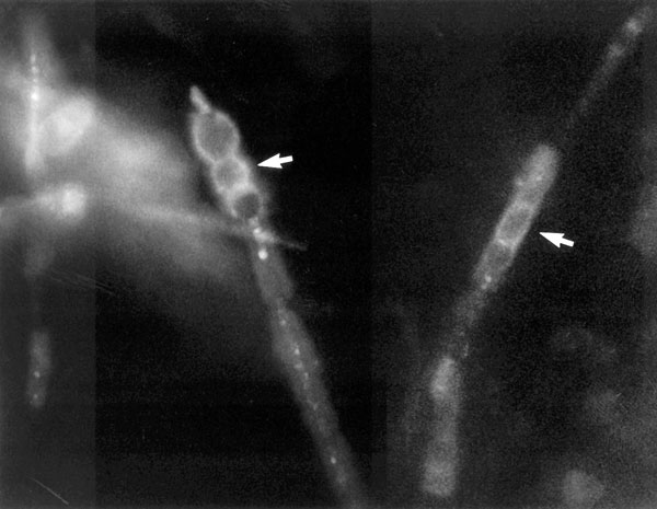
Figure 1. Coccidioides immitis hyphae dislodged from the tip of the ventriculo-peritoneal catheter tip, fixed in formalin and stained with Calcofluor. Stained barrel-shaped arthroconidia (arrows) are seen, along with empty cells (x400). The upper left image shows a section from the tip of the shunt tubing stained with SYTOX Green nucleic acid stain and examined by scanning confocal microscopy with argon-ion laser light source. This specific staining for nucleic acids clearly shows the presence of a biofilm and some 4- to 6-μm cells. The upper right image shows an unstained, transmitted light microscopic image of the same area of the edge of the tubing. The bottom right image shows a recombined image with the nucleic acid stain colocalized with the transmitted light image. The recombined image shows that a substantial (~30 μm) biofilm composed of 4- to 6-μm cells has colonized the “scalloped” surface of this tubing. (x630 total magnification mosaic)