Volume 9, Number 10—October 2003
Dispatch
West Nile Virus Encephalitis and Myocarditis in Wolf and Dog
Figure 1
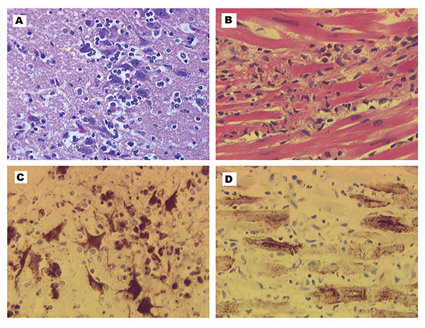
Figure 1. Encephalitis and myocarditis in two West Nile virus–infected canids. A, histopathology of the cerebrum of the wolf. Focus of lymphocyte infiltration and necrosis with mild gliosis; arrow indicates necrotic neuron. Hematoxylin-eosin staining. B, histopathology of the heart of the dog. Focus of leukocytic infiltration, mostly lymphocytes, and myocardial cell degeneration and necrosis. Hematoxylin-eosin staining. C, West Nile virus immunohistochemistry of the cerebrum of the wolf. Intense labeling of neurons in focus of inflammation. Some labeling is also in the glial cells or lymphocytes. Immunoperoxidase with hematoxylin counterstain. D, West Nile virus immunohistochemistry of the heart of dog. Intense labeling of most myocardial cells in the field. Immunoimmunoperoxidase with hematoxylin counterstain.