Volume 13, Number 8—August 2007
Research
Classic Scrapie in Sheep with the ARR/ARR Prion Genotype in Germany and France
Martin H. Groschup*1 , Caroline Lacroux†1, Anne Buschmann*, Gesine Lühken‡, Jacinthe Mathey†, Martin Eiden*, Séverine Lugan†, Christine Hoffmann*, Juan Carlos Espinosa§, Thierry G.M. Baron¶, Juan Maria Torres§, Georg Erhardt‡, and Olivier Andreoletti†
Figure 3
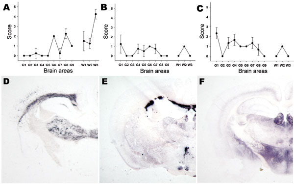
Figure 3. Lesion profiling (A, B, C) and paraffin-embedded tissue blot characterization of prion protein (PrPSc) deposition at thalamic level (D, E, F). Tests were performed by using formalin-fixed brain from Tg338 mice (expressing the VRQ PrP ovine variant) inoculated with (A, D) ARR/ARR atypical case (B, E) bovine spongiform encephalopathy (BSE) brain from an ARR/ARR sheep (intracerebral inoculation), and (C, F) case S83. Each lesion profile was carried out by using 6 animals. Detection of PrPSc was achieved by using the monoclonal antibody Sha31.
Page created: June 30, 2010
Page updated: June 30, 2010
Page reviewed: June 30, 2010
The conclusions, findings, and opinions expressed by authors contributing to this journal do not necessarily reflect the official position of the U.S. Department of Health and Human Services, the Public Health Service, the Centers for Disease Control and Prevention, or the authors' affiliated institutions. Use of trade names is for identification only and does not imply endorsement by any of the groups named above.