Skip directly to site content Skip directly to page options Skip directly to A-Z link Skip directly to A-Z link Skip directly to A-Z link
Volume 13, Number 9—September 2007
Dispatch

Coronavirus Antibodies in African Bat Species

Marcel A. Müller*Comments to Author , Janusz T. Paweska†, Patricia A. Leman†, Sung Sup Park‡, Klaus Grywna‡, Alan Kemp†, Leo Braack§, Karen Sonnenberg¶, Matthias Niedrig*, and Robert Swanepoel†
Author affiliations: *Robert Koch-Institut, Berlin, Germany; †National Institute for Communicable Diseases, Sandringham, Republic of South Africa; ‡Bernhard Nocht Institute for Tropical Medicine, Hamburg, Germany; §Conservation International, Cape Town, Republic of South Africa; ¶EUROIMMUN AG, Lübeck, Germany;

Main Article

Figure 2

Results of indirect immunofluorescence (IF) test with Vero E6 cells infected with severe acute respiratory syndrome–associated coronavirus (SARS-CoV). The SARS-CoV diagnostic IIFT kit (EUROIMMUN AG, Lübeck, Germany) was used with minor modifications: bat and reference human serum specimens were diluted 1:100 (found to be the optimal dilution for bat sera) in sample buffer, and secondary detection was performed with goat-antibat immunoglobulin (Ig) (Bethyl, Montgomery, AL, USA) followed by fluorescein isothiocyanate (FITC)–labeled donkey-antigoat Ig (Dianova, Hamburg, Germany) (A–F) or FITC-labeled goat-antihuman Ig (G–I). Frames A–D, SARS-CoV ELISA–positive bat serum specimens 2, 17, 26, 31; E–F, ELISA-negative bat serum specimens 38 (showing unspecific signals) and 306; G–H, SARS-CoV–positive human control serum specimens A and B; I, negative human serum C. All photographs were taken at equivalent microscope settings. Scale bars represent 20 μm.

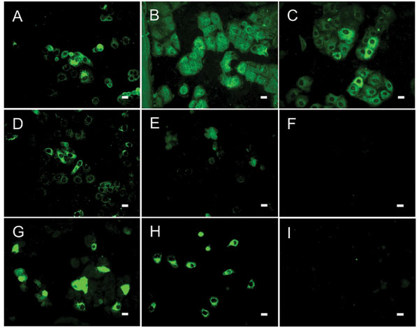
Figure 2. Results of indirect immunofluorescence (IF) test with Vero E6 cells infected with severe acute respiratory syndrome–associated coronavirus (SARS-CoV). The SARS-CoV diagnostic IIFT kit (EUROIMMUN AG, Lübeck, Germany) was used with minor modifications: bat and reference human serum specimens were diluted 1:100 (found to be the optimal dilution for bat sera) in sample buffer, and secondary detection was performed with goat-antibat immunoglobulin (Ig) (Bethyl, Montgomery, AL, USA) followed by fluorescein isothiocyanate (FITC)–labeled donkey-antigoat Ig (Dianova, Hamburg, Germany) (A–F) or FITC-labeled goat-antihuman Ig (G–I). Frames A–D, SARS-CoV ELISA–positive bat serum specimens 2, 17, 26, 31; E–F, ELISA-negative bat serum specimens 38 (showing unspecific signals) and 306; G–H, SARS-CoV–positive human control serum specimens A and B; I, negative human serum C. All photographs were taken at equivalent microscope settings. Scale bars represent 20 μm.

Main Article

Page created: July 01, 2010
Page updated: July 01, 2010
Page reviewed: July 01, 2010
The conclusions, findings, and opinions expressed by authors contributing to this journal do not necessarily reflect the official position of the U.S. Department of Health and Human Services, the Public Health Service, the Centers for Disease Control and Prevention, or the authors' affiliated institutions. Use of trade names is for identification only and does not imply endorsement by any of the groups named above.
file_external