Volume 14, Number 11—November 2008
Letter
Rickettsia aeschlimannii Infection, Algeria
Figure
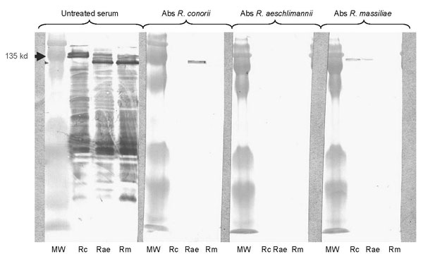
Figure. Western blot assay (WB) and cross-adsorption studies in serum of a patient with rickettsiosis in Algeria. Immunofluorescent assay showed raised levels of immunoglobulin (Ig) G/M at the same titer (2,048/32) against Rickettsia conorii, R. aeschlimannii, and R. massiliae. Lanes Rc, Rae, and Rm: WB assay using R. conorii, R. aeschlimannii, and R. massiliae antigens, respectively. MW, molecular weights are indicated on the left. Untreated serum, late serum samples tested by WB. When adsorption is performed with R. aeschlimannii antigens, homologous and heterologous antibodies disappear, but when it is performed with R. conorii antigens and R. massiliae, homologous antibodies disappear but heterologous antibodies persist. This result indicates that antibodies are specifically directed against R. aeschlimannii. Abs, absorbed.