Volume 14, Number 3—March 2008
Dispatch
Hemagglutinating Encephalomyelitis Coronavirus Infection in Pigs, Argentina
Maria A. Quiroga* , Javier Cappuccio*, Pablo E. Piñeyro*, Walter Basso*, Gastón Moré*, Mariana Kienast†, Sergio Schonfeld‡, José L. Cáncer‡, Sandra Arauz*, María E. Pintos*, Mariana Nanni†, Mariana Machuca*, Norio Hirano§, and Carlos J. Perfumo*
Figure 1
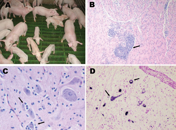
Figure 1. A) Nursery piglets showing clinical signs compatible with porcine hemagglutinating encephalomyelitis coronavirus (PHE-CoV). Nonaffected pigs of the same age are also shown. B) Muscle layer of stomach from affected piglet showing perivascular cuffing (arrow); hematoxylin-eosin stain, magnification ×100. C) Brainstem from affected piglet showing satellitosis (arrows) and gliosis; hematoxylin-eosin stain, magnification x400. D) Brainstem from affected piglet showing positive label of neuron perikarion (arrows); nitroblue-tetrazolium imunohistochemical stain, magnification x400.
Page created: July 07, 2010
Page updated: July 07, 2010
Page reviewed: July 07, 2010
The conclusions, findings, and opinions expressed by authors contributing to this journal do not necessarily reflect the official position of the U.S. Department of Health and Human Services, the Public Health Service, the Centers for Disease Control and Prevention, or the authors' affiliated institutions. Use of trade names is for identification only and does not imply endorsement by any of the groups named above.