Volume 15, Number 10—October 2009
Letter
Rhombencephalitis and Coxsackievirus A16
Kazuna Goto, Masafumi Sanefuji , Koichi Kusuhara1, Yorihiro Nishimura, Hiroyuki Shimizu, Ryutaro Kira, Hiroyuki Torisu, and Toshiro Hara
Figure
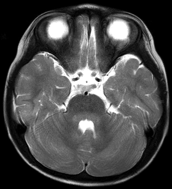
Figure. Axial T2-weighted slice of brain by magnetic resonance imaging, showing hyperintensity lesions in the pons and cerebellum around the fourth ventricle.
Page created: December 08, 2010
Page updated: December 08, 2010
Page reviewed: December 08, 2010
The conclusions, findings, and opinions expressed by authors contributing to this journal do not necessarily reflect the official position of the U.S. Department of Health and Human Services, the Public Health Service, the Centers for Disease Control and Prevention, or the authors' affiliated institutions. Use of trade names is for identification only and does not imply endorsement by any of the groups named above.