Volume 15, Number 11—November 2009
Dispatch
Fatal Case of Enterovirus 71 Infection, France, 2007
Figure 2
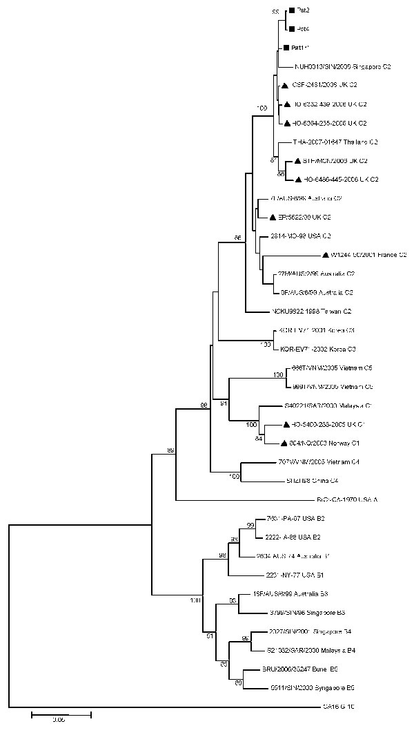
Figure 2. Phylogenetic relationships between 3 French strains and 34 worldwide enterovirus 71 GenBank-selected strains based on alignment of complete viral protein (VP) 1 coding sequences. The prototype coxsackievirus A16 (CoxA16-G10) was used as the outgroup virus. The phylogenetic tree was constructed by the neighbor-joining method by using MEGA4 (www.megasoftware.net). Bootstrap values (>70%) derived from 1,000 samplings are shown at the nodes of the tree. Phylogenetic separation of C2 isolates appear in accordance with time and place of isolation. Isolates from this study are indicated by black squares (the strain isolated from the 17-month-old boy with fatal pulmonary edema is shown in boldface with **), and the other European circulating strains by black triangles. Branch lengths are drawn to scale. Scale bar indicates 5% of nucleotide sequence divergence.