Volume 15, Number 12—December 2009
Dispatch
Echinococcus vogeli Infection in a Hunter, French Guiana
Jenny Knapp1, Mircea Chirica, Christine Simonnet, Frederic Grenouillet, Jean-Mathieu Bart, Yasuhito Sako, Sonoyo Itoh, Minoru Nakao, Akira Ito1, and Laurence Millon1
Figure 1
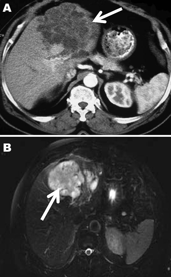
Figure 1. Computed tomography (A) and magnetic resonance (B) images of the liver of a 72-year-old man from French Guiana with polycystic echinococcosis affecting the left side of the liver. White arrows indicate the multicystic liver lesion.
Page created: December 09, 2010
Page updated: December 09, 2010
Page reviewed: December 09, 2010
The conclusions, findings, and opinions expressed by authors contributing to this journal do not necessarily reflect the official position of the U.S. Department of Health and Human Services, the Public Health Service, the Centers for Disease Control and Prevention, or the authors' affiliated institutions. Use of trade names is for identification only and does not imply endorsement by any of the groups named above.