Skip directly to site content Skip directly to page options Skip directly to A-Z link Skip directly to A-Z link Skip directly to A-Z link
Volume 15, Number 8—August 2009
Research

Transgenic Mice Expressing Porcine Prion Protein Resistant to Classical Scrapie but Susceptible to Sheep Bovine Spongiform Encephalopathy and Atypical Scrapie

Juan-Carlos Espinosa1, María-Eugenia Herva1, Olivier Andréoletti, Danielle Padilla, Caroline Lacroux, Hervé Cassard, Isabelle Lantier, Joaquin Castilla, and Juan-María TorresComments to Author 
Author affiliations: Centro de Investigación en Sanidad Animal, Madrid, Spain (J.-C. Espinosa, M.-E. Herva, D. Padilla, J. Castilla, J.-M. Torres); École Nationale Vétérinaire de Toulouse, Toulouse, France (O. Andréoletti, C. Lacroux, H. Cassard); Centre Institut National de la Recherche Agronomique de Tours, Nouzilly, France (I. Lantier)

Main Article

Figure 4

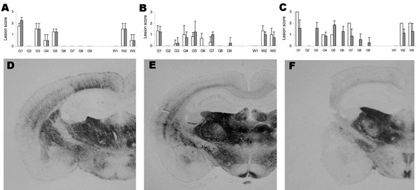
Lesion profiles and regional distributions of atypical proteinase K–resistant prion protein (PrPres) in the brain of porcine PrP transgenic mice infected, either in 1st passage (white column) or in 2nd passage (black column) with cattle bovine spongiform encephalopathy (BSE) (panels A and D), sheep BSE (panels B and E), or atypical scrapie (panels C and F) agents. A–C) Lesion scoring of 9 areas of gray matter (G) and white matter (W) in mice brains: dorsal medulla (G1), cerebellar cortex (G2), s

Figure 4. Lesion profiles and regional distributions of atypical proteinase K–resistant prion protein (PrPres) in the brain of porcine PrP transgenic mice infected, either in 1st passage (white column) or in 2nd passage (black column) with cattle bovine spongiform encephalopathy (BSE) (panels A and D), sheep BSE (panels B and E), or atypical scrapie (panels C and F) agents. A–C) Lesion scoring of 9 areas of gray matter (G) and white matter (W) in mice brains: dorsal medulla (G1), cerebellar cortex (G2), superior colliculus (G3), hypothalamus (G4), medial thalamus (G5), hippocampus (G6), septum (G7), medial cerebral cortex at the level of the thalamus (G8) and at the level of the septum (G9), cerebellum (W1), mesencephalic tegmentum (W2) and pyramidal tract (W3). Error bars indicate SE. D–F) Histoblots of representative coronal sections at the level of the hippocampus.

Main Article

1These authors contributed equally to this article.
Page created: October 04, 2010
Page updated: October 04, 2010
Page reviewed: October 04, 2010
The conclusions, findings, and opinions expressed by authors contributing to this journal do not necessarily reflect the official position of the U.S. Department of Health and Human Services, the Public Health Service, the Centers for Disease Control and Prevention, or the authors' affiliated institutions. Use of trade names is for identification only and does not imply endorsement by any of the groups named above.
file_external