Volume 15, Number 8—August 2009
Dispatch
Lobomycosis in Man and Lobomycosis-like Disease in Bottlenose Dolphin, Venezuela
Luis Bermudez, Marie-Françoise Van Bressem, Oscar Reyes-Jaimes, Alejandro J. Sayegh, and Alberto Enrique Paniz-Mondolfi
Figure 1
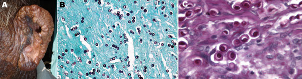
Figure 1. A) Multiple, confluent, keloid-like, hyperchromic nodules with flat shiny surfaces involving the entire free border, posterior aspect, and lobule of the left ear of a fisherman, Venezuela. B) Numerous Lacazia loboi tissue-phase organisms within the stroma. Note the typical chain pattern showing simple gemation budding (Gomori-Grocott stain, magnification ×100). C) Yeast cells showing typical double refraction of the membrane and protoplasmic bodies within cells (periodic acid–Schiff stain, magnification ×600).
Page created: November 01, 2010
Page updated: November 01, 2010
Page reviewed: November 01, 2010
The conclusions, findings, and opinions expressed by authors contributing to this journal do not necessarily reflect the official position of the U.S. Department of Health and Human Services, the Public Health Service, the Centers for Disease Control and Prevention, or the authors' affiliated institutions. Use of trade names is for identification only and does not imply endorsement by any of the groups named above.