Volume 16, Number 3—March 2010
Research
Use of Avian Bornavirus Isolates to Induce Proventricular Dilatation Disease in Conures
Patricia Gray, Sharman Hoppes, Paulette Suchodolski, Negin Mirhosseini, Susan Payne, Itamar Villanueva, H.L Shivaprasad, Kirsi S. Honkavuori, Thomas Briese, Sanjay M. Reddy, and Ian Tizard
Figure 1
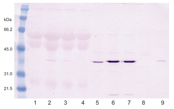
Figure 1. Western blot of infected duck embryonic fibroblasts (DEFs) showing avian bornavirus N-protein during culture. Lanes 1–4 are supernatant fluids. Lane I is from an African gray parrot (AG5). Lanes 2 and 3 are from a yellow-collared macaw (M24). Lane 4 is from uninfected DEFs. Lanes 5–8 are sonicated cell extracts. Lane 5 from AG5; 6 and 7 from M24; and Lane 8 from uninfected DEFs. Lane 9 is an infected brain control. The virus is strongly cell associated.
Page created: December 14, 2010
Page updated: December 14, 2010
Page reviewed: December 14, 2010
The conclusions, findings, and opinions expressed by authors contributing to this journal do not necessarily reflect the official position of the U.S. Department of Health and Human Services, the Public Health Service, the Centers for Disease Control and Prevention, or the authors' affiliated institutions. Use of trade names is for identification only and does not imply endorsement by any of the groups named above.