Volume 16, Number 3—March 2010
Dispatch
Influenza A Pandemic (H1N1) 2009 Virus Infection in Domestic Cat
Figure

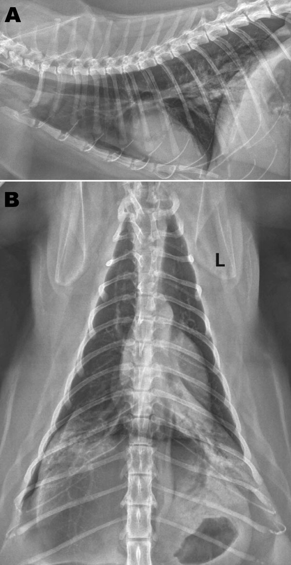
Figure. Radiographs of the thorax of a cat with confirmed influenza A pandemic (H1N1) 2009 virus infection. A) Right lateral view; B) dorsoventral view. Asymmetric soft tissue opacities are evident in the right and left caudal lung lobes. An alveolar pattern, composed of air bronchograms with border-effaced (indistinct) adjacent pulmonary vessels, is most pronounced in the left caudal lobe. A small gas lucency in the pleural space appears in the right caudal and dorsal thoracic cavity. An endotracheal tube is visible at the thoracic inlet on the lateral view in this moderately obese cat. L, left.
Page created: December 14, 2010
Page updated: December 14, 2010
Page reviewed: December 14, 2010
The conclusions, findings, and opinions expressed by authors contributing to this journal do not necessarily reflect the official position of the U.S. Department of Health and Human Services, the Public Health Service, the Centers for Disease Control and Prevention, or the authors' affiliated institutions. Use of trade names is for identification only and does not imply endorsement by any of the groups named above.