Volume 16, Number 5—May 2010
Dispatch
Adenovirus 36 DNA in Adipose Tissue of Patient with Unusual Visceral Obesity
Behrouz Salehian, Stephen J. Forman, Fouad R. Kandeel, Denise E. Bruner, Jia He, and Richard L. Atkinson
Figure 1
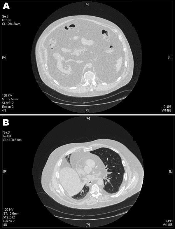
Figure 1. Computed tomography scans of the patient, showing marked visceral adipose tissue in the abdomen (A) and thorax (B). Diffuse intrabadominal, retroperitoneal lipomatosis, and herniation of the mediastinum can be seen through the esophageal hiatus. Intrapericardial adipose infiltration and adipose tissue bilaterally are seen within the pleura.
Page created: December 23, 2010
Page updated: December 23, 2010
Page reviewed: December 23, 2010
The conclusions, findings, and opinions expressed by authors contributing to this journal do not necessarily reflect the official position of the U.S. Department of Health and Human Services, the Public Health Service, the Centers for Disease Control and Prevention, or the authors' affiliated institutions. Use of trade names is for identification only and does not imply endorsement by any of the groups named above.