Volume 16, Number 8—August 2010
Dispatch
Amblyomma imitator Ticks as Vectors of Rickettsia rickettsii, Mexico
Karla A. Oliveira1, Adriano Pinter2, Aaron Medina-Sanchez3, Venkata D. Boppana, Stephen K. Wikel, Tais B. Saito, Thomas Shelite, Lucas S. Blanton, Vsevolod Popov, Pete D. Teel4, David H. Walker, Marcio A.M. Galvao5, Claudio Mafra1, and Donald H. Bouyer
Figure
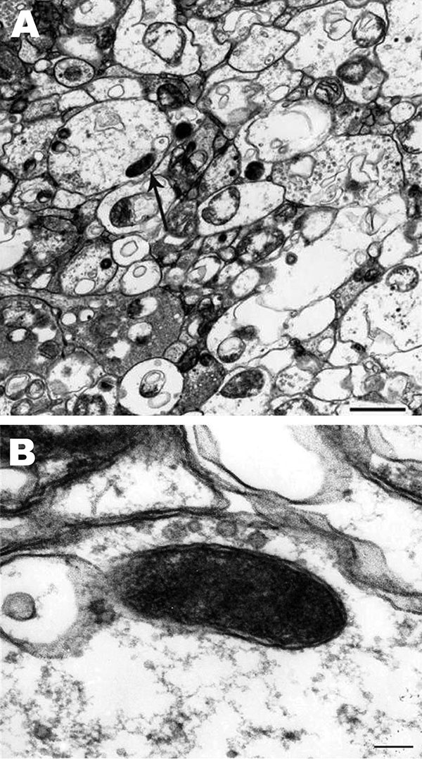
Figure. Rickettsia rickettsii (arrow) in a midgut cell of an Amblyomma imitator tick (A). The trilaminar cell wall is separated from the cell membrane by the periplasmic space (B). Scale bars = 0.1 µm.
1Current affiliation: Federal University of Vicosa, Vicosa, Brazil.
2Current affiliation: Superintendency of Control of Endemic Diseases, São Paulo, Brazil.
3Current affiliation: Hospital and Clinic Oca, Monterrey, Nuevo Leon, Mexico.
4Current affiliation: Texas A&M University, College Station, Texas, USA.
5Current affiliation: Federal University of Ouro Preto, Ouro Preto, Brazil.
Page created: March 30, 2011
Page updated: March 30, 2011
Page reviewed: March 30, 2011
The conclusions, findings, and opinions expressed by authors contributing to this journal do not necessarily reflect the official position of the U.S. Department of Health and Human Services, the Public Health Service, the Centers for Disease Control and Prevention, or the authors' affiliated institutions. Use of trade names is for identification only and does not imply endorsement by any of the groups named above.