Volume 6, Number 2—April 2000
Dispatch
Chlamydia pneumoniae Infection in a Breeding Colony of African Clawed Frogs (Xenopus tropicalis)
Kurt D. Reed* , George R. Ruth†, Jeanine A. Meyer*, and Sanjay K. Shukla*
Figure
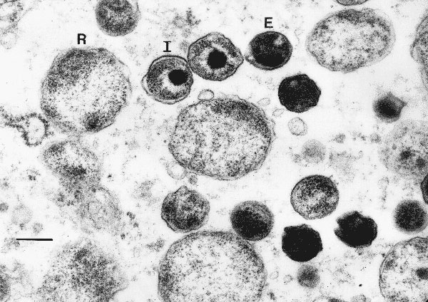
Figure. Transmission electron micrograph of chlamydial particles in liver from a captive African clawed frog (Xenopus tropicalis). Note the reticulate bodies (R), intermediate bodies (I), and highly condensed elementary bodies (E). Bar, 270 nm.
Page created: December 16, 2010
Page updated: December 16, 2010
Page reviewed: December 16, 2010
The conclusions, findings, and opinions expressed by authors contributing to this journal do not necessarily reflect the official position of the U.S. Department of Health and Human Services, the Public Health Service, the Centers for Disease Control and Prevention, or the authors' affiliated institutions. Use of trade names is for identification only and does not imply endorsement by any of the groups named above.