Volume 10, Number 12—December 2004
Research
Origin of the Amphibian Chytrid Fungus
Ché Weldon* , Louis H. du Preez*, Alex D. Hyatt†, Reinhold Muller‡, and Rick Speare‡
Figure 1
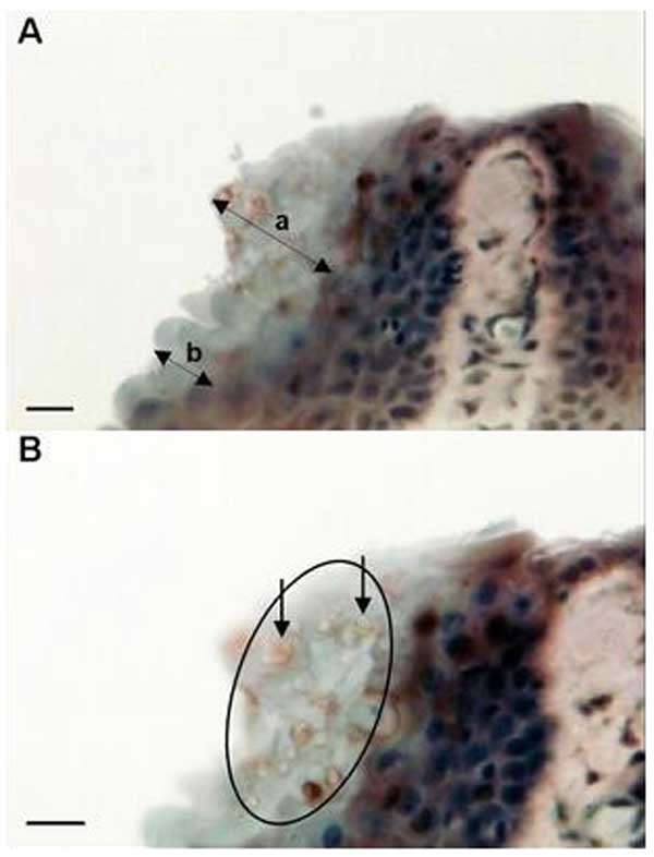
Figure 1. Micrographs of immunoperoxidase stained sections through the interdigital webbing of Xenopus gilli, showing the morphologic features and size of zoosporangia consistent with Batrachochytrium dendrobatidis. A) Arrow a indicates localized hyperplastic epidermal response; arrow b indicates an uninfected region of the epidermis. B) Arrows indicate two zoosporangia with internal septa. Circle indicates location of the infection in the stratum corneum. Bar, 10 μm.
Page created: April 14, 2011
Page updated: April 14, 2011
Page reviewed: April 14, 2011
The conclusions, findings, and opinions expressed by authors contributing to this journal do not necessarily reflect the official position of the U.S. Department of Health and Human Services, the Public Health Service, the Centers for Disease Control and Prevention, or the authors' affiliated institutions. Use of trade names is for identification only and does not imply endorsement by any of the groups named above.