Skip directly to site content Skip directly to page options Skip directly to A-Z link Skip directly to A-Z link Skip directly to A-Z link
Volume 10, Number 7—July 2004
Dispatch

Mice Susceptible to SARS Coronavirus

David E. Wentworth*Comments to Author , Laura Gillim-Ross*, Noel Espina*, and Kristen A. Bernard*†
Author affiliations: *New York Department of Health, Albany, New York, USA; †State University of New York, Albany, New York, USA

Main Article

Figure 1

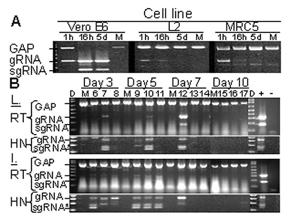
Replication-specific multiplex reverse transcriptase–polymerase chain reaction (RT-PCR) assay shows severe acute respiratory syndrome–associated coronavirus (SARS-CoV) replicated in the lungs and intestines of mice. A. Vero E6, murine fibroblast (L2), and human lung fibroblasts (MRC5) were inoculated with SARS-CoV at an MOI of ≈ 0.001 or were mock-inoculated (M). G3PDH, SARS-CoV gRNA, and sgRNA were amplified by multiplex RT-PCR from total RNA extracted at 1 h, 16 h, or 5 days after inoculation.

Figure 1. Replication-specific multiplex reverse transcriptase–polymerase chain reaction (RT-PCR) assay shows severe acute respiratory syndrome–associated coronavirus (SARS-CoV) replicated in the lungs and intestines of mice. A. Vero E6, murine fibroblast (L2), and human lung fibroblasts (MRC5) were inoculated with SARS-CoV at an MOI of ≈ 0.001 or were mock-inoculated (M). G3PDH, SARS-CoV gRNA, and sgRNA were amplified by multiplex RT-PCR from total RNA extracted at 1 h, 16 h, or 5 days after inoculation. Amplicons were visualized by ethidium bromide staining after electrophoresis; negative images are shown. B. Mice were inoculated with 2x10⁵ 50% tissue culture infective dose of SARS-CoV (lanes labeled 6–17) or were mock-inoculated (M) and euthanized after 3, 5, 7, or 10 days. G3PDH, SARS-CoV gRNA, and sgRNA were amplified by multiplex RT-PCR from total RNA extracted from the lung (L) and intestine (I) harvested at various time points. Heminested PCR (HN) was used to amplify gRNA and sgRNA from RT-PCR reactions. Positive and negative controls for PCR reactions are indicated by + and –, respectively. D indicates DNA marker ladder. *The doublet observed in HN-PCR reactions results from residual primers used in the primary amplification reaction.

Main Article

Page created: January 27, 2011
Page updated: January 27, 2011
Page reviewed: January 27, 2011
The conclusions, findings, and opinions expressed by authors contributing to this journal do not necessarily reflect the official position of the U.S. Department of Health and Human Services, the Public Health Service, the Centers for Disease Control and Prevention, or the authors' affiliated institutions. Use of trade names is for identification only and does not imply endorsement by any of the groups named above.
file_external