Volume 11, Number 1—January 2005
Letter
Disseminated Coccidioidomycosis
Cheng-Yi Wang*, Jih-Shuin Jerng*, Jen-Chung Ko†, Ming-Feng Lin†, Cheng-Hsiang Hsiao*, Li-Na Lee*, Po-Ren Hsueh* , and Sow-Hsong Kuo*
Figure
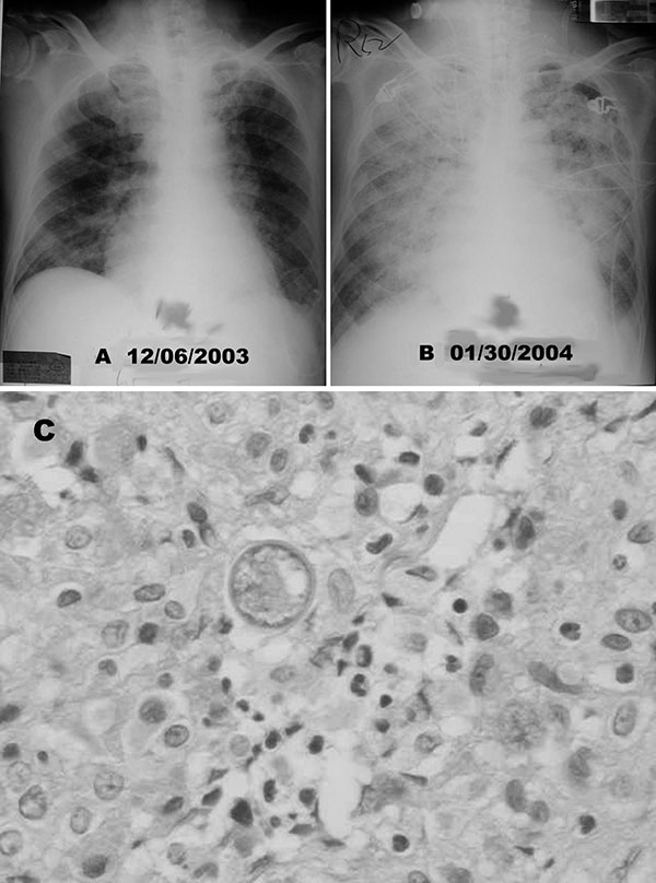
Figure. A) Chest radiograph shows diffuse nodular lesions in both lungs. B) Chest radiographic scan taken 2 months later shows coalescence of nodular shadows and almost complete white-out of bilateral lung fields. C) Hematoxylin and eosin staining of the wound specimen from pleural biopsy site showed spherules of Coccidioides immitis and chronic necrotizing granulomatous inflammation (400x).
Page created: April 15, 2011
Page updated: April 15, 2011
Page reviewed: April 15, 2011
The conclusions, findings, and opinions expressed by authors contributing to this journal do not necessarily reflect the official position of the U.S. Department of Health and Human Services, the Public Health Service, the Centers for Disease Control and Prevention, or the authors' affiliated institutions. Use of trade names is for identification only and does not imply endorsement by any of the groups named above.