Volume 11, Number 11—November 2005
Synopsis
Coltiviruses and Seadornaviruses in North America, Europe, and Asia
Houssam Attoui* , Fauziah Mohd Jaafar*, Philippe de Micco*, and Xavier de Lamballerie*
Figure 1
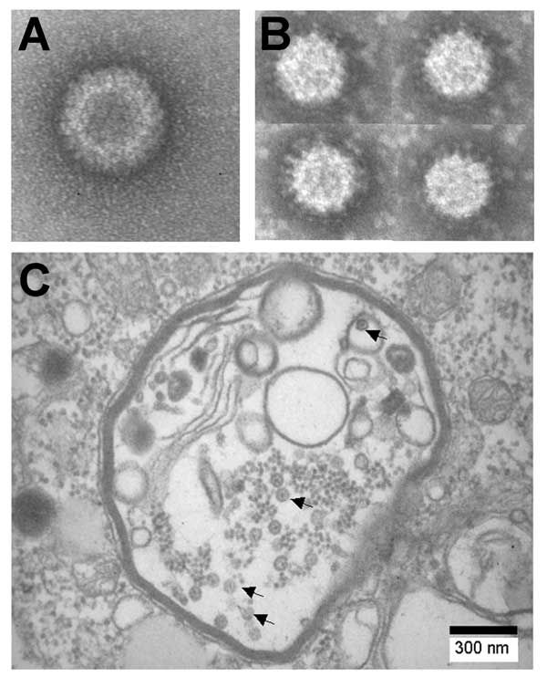
Figure 1. Negative contrast electron micrographs of A) Colorado tick fever virus and B) Banna virus (BAV). C) Thin section of BAV-infected C6/36 cells showing viral particles (arrows) in vacuolelike structures.
Page created: February 17, 2012
Page updated: February 17, 2012
Page reviewed: February 17, 2012
The conclusions, findings, and opinions expressed by authors contributing to this journal do not necessarily reflect the official position of the U.S. Department of Health and Human Services, the Public Health Service, the Centers for Disease Control and Prevention, or the authors' affiliated institutions. Use of trade names is for identification only and does not imply endorsement by any of the groups named above.