Volume 11, Number 12—December 2005
Perspective
Human Granulocytic Anaplasmosis and Anaplasma phagocytophilum
J. Stephen Dumler* , Kyoung-Seong Choi*, Jose Carlos Garcia-Garcia*, Nicole S. Barat*, Diana G. Scorpio*, Justin W. Garyu*, Dennis J. Grab*, and Johan S. Bakken†‡
Figure 1
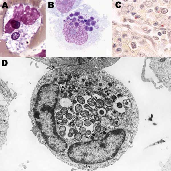
Figure 1. Anaplasma phagocytophilum in human peripheral blood band neutrophil (A. Wright stain, original magnification ×1,000), in THP-1 myelomonocytic cell culture (B, LeukoStat stain, original magnification, ×400), in neutrophils infiltrating human spleen (C, immunohistochemistry with hematoxylin counterstain; original magnification ×100), and ultrastructure by transmission electron microscopy in HL-60 cell culture (D; courtesy of V. Popov; original magnification ×21,960).
Page created: February 02, 2012
Page updated: February 02, 2012
Page reviewed: February 02, 2012
The conclusions, findings, and opinions expressed by authors contributing to this journal do not necessarily reflect the official position of the U.S. Department of Health and Human Services, the Public Health Service, the Centers for Disease Control and Prevention, or the authors' affiliated institutions. Use of trade names is for identification only and does not imply endorsement by any of the groups named above.