Human Disease from Influenza A (H5N1), Thailand, 2004
Tawee Chotpitayasunondh*, Kumnuan Ungchusak†, Wanna Hanshaoworakul†, Supamit Chunsuthiwat†, Pathom Sawanpanyalert†, Rungruen Kijphati†, Sorasak Lochindarat*, Panida Srisan*, Pongsan Suwan†, Yutthasak Osotthanakorn†, Tanakorn Anantasetagoon†, Supornchai Kanjanawasri†, Sureeporn Tanupattarachai†, Jiranun Weerakul†, Ruangsri Chaiwirattana†, Monthira Maneerattanaporn†, Rapol Poolsavatkitikool†, Kulkunya Chokephaibulkit‡, Anucha Apisarnthanarak§, and Scott F. Dowell¶

Author affiliations: *Queen Sirikit National Institute of Child Health, Bangkok, Thailand; †Ministry of Public Health, Nonthaburi, Thailand; ‡Siriraj Hospital, Bangkok, Thailand; §Thammasat University Hospital, Bangkok, Thailand; ¶International Emerging Infections Program, Nonthaburi, Thailand
Main Article
Figure 5
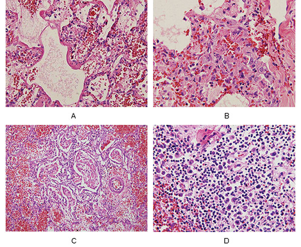
Figure 5. Pathologic findings from a patient (number 6) with confirmed influenza A (H5N1) infection. All slides are stained with hematoxylin and eosin, shown at 40x objective. Panel A shows hyaline membrane formation lining the alveolar spaces of the lung and vascular congestion with a few infiltrating lymphocytes in the interstitial areas. Reactive fibroblasts are also present. Panel B is an area of lung with proliferating reactive fibroblasts within the interstitial areas. Few lymphocytes are seen, and no viral intranuclear inclusions are visible. Panel C shows fibrinous exudates filling the alveolar spaces, with organizing formation and few hyaline membranes. The surrounding alveolar spaces contain hemorrhage. Panel D is from a section of spleen, showing numerous atypical lymphoid cells scattered around the white pulp. No viral intranuclear inclusions are seen.
Main Article
Page created: April 28, 2011
Page updated: April 28, 2011
Page reviewed: April 28, 2011
The conclusions, findings, and opinions expressed by authors contributing to this journal do not necessarily reflect the official position of the U.S. Department of Health and Human Services, the Public Health Service, the Centers for Disease Control and Prevention, or the authors' affiliated institutions. Use of trade names is for identification only and does not imply endorsement by any of the groups named above.