Volume 11, Number 3—March 2005
Dispatch
Human Metapneumovirus RNA in Encephalitis Patient
Oliver Schildgen*1 , Thomas Glatzel*1, Tilman Geikowski*, Bärbel Scheibner*, Arne Simon*, Lutz Bindl*, Mark Born*, Sergei Viazov†, Anja Wilkesmann*, Gisela Knöpfle*, Michael Roggendorf†, and Bertfried Matz*
Figure 1
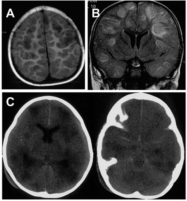
Figure 1. . Axial T1-weighted magnetic resonance imaging (MRI) scan (A) and coronal fluid attenuated inversion recovery (FLAIR) (B) show multifocal, mainly cortical and subcortical lesions of high signal intensity, which are most probably caused by multifocal encephalitis. C) Nonenhanced axial computed tomographic (CT) scan performed 2 days after the MRI shows multiple, hypodense lesions and signs of general edema. Additionally, it shows a hyperdense arachnoid collection that was not yet visible on the MRI 2 days before (panels A and B).
Page created: April 25, 2012
Page updated: April 25, 2012
Page reviewed: April 25, 2012
The conclusions, findings, and opinions expressed by authors contributing to this journal do not necessarily reflect the official position of the U.S. Department of Health and Human Services, the Public Health Service, the Centers for Disease Control and Prevention, or the authors' affiliated institutions. Use of trade names is for identification only and does not imply endorsement by any of the groups named above.