Volume 11, Number 5—May 2005
Research
Adenovirus Type 7 Peptide Diversity during Outbreak, Korea, 1995–2000
Eun Hwa Choi*†, Hee Sup Kim‡, Byung Wook Eun*§, Beyong Il Kim*, Jung Yeon Choi*, Hoan Jong Lee*§ , and Toshiki Inada¶
Figure 1
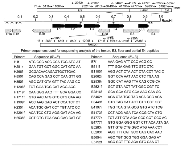
Figure 1. . Schematic representation of the restriction mapping sites of adenovirus type 7d (Ad7d) and Ad7l by BamHI, the primer sequences, and their location for the sequencing analysis of hexon, E3, fiber, and E4 open reading frame (ORF) 6/7 peptides. Each restriction site by BamHI is indicated as (↑). The restriction site at 0.93 map units shown by (↑*) is lost in strains with genome type Ad7l and present in Ad7d. Figure of each primer represents H, hexon; E, E3, fiber; and E4 ORF 6/7 peptides, the position of the first nucleotide at the 5´ end; f, forward; and r, reverse. The numbering of nucleotides is based on that of strain 383, which was isolated in Japan in 1992 (AF053086 for hexon and AF104383 for E3, fiber, and E4).
Page created: April 24, 2012
Page updated: April 24, 2012
Page reviewed: April 24, 2012
The conclusions, findings, and opinions expressed by authors contributing to this journal do not necessarily reflect the official position of the U.S. Department of Health and Human Services, the Public Health Service, the Centers for Disease Control and Prevention, or the authors' affiliated institutions. Use of trade names is for identification only and does not imply endorsement by any of the groups named above.