Volume 12, Number 3—March 2006
Research
Aspergillus ustus Infections among Transplant Recipients
Cite This Article
Citation for Media
Abstract
Aspergillus ustus is a mold that rarely infects humans; only 15 systemic cases have been reported. We report the first outbreak of invasive infection caused by A. ustus among hematopoietic stem cell transplant (HSCT) recipients. Six patients with infections were identified; 3 infections each occurred in both 2001 and 2003. Molecular typing by using randomly amplified polymorphic DNA (RAPD) and antifungal drug susceptibility testing were performed on clinical and environmental isolates recovered from our hospital from 1999 to 2003. The highest overall attack rate in HSCT patients was 1.6%. The overall death rate was 50%, and death occurred within 8 days after diagnostic culture collection. Clinical isolates exhibited decreased susceptibility to antifungal drugs, especially azoles. RAPD and phylogenetic analysis showed genetic similarity between isolates from different patients. Based on the clustering of cases in space and time and molecular data, common-source acquisition of this unusual drug-resistant species is possible.
Invasive aspergillosis (IA) has become a devastating opportunistic fungal infection among immunocompromised hosts, with a 357% increase in death rates reported in the United States from 1980 to 1997 (1). The most common cause of IA is Aspergillus fumigatus (2). However, in recent years, IA has been increasingly caused by non-fumigatus Aspergillus species. For example, at the Fred Hutchinson Cancer Research Center in Seattle, the proportion of infections caused by non-fumigatus Aspergillus species increased during the latter 1990s. Most of these infections were caused by A. flavus, A. nidulans, A. terreus, and A. niger (3).
Aspergillus ustus is a group of filamentous hyalohyphomycetes consisting of 5 species: A. ustus, A. puniceus, A. panamensis, A. conjunctus, and A. deflectus. Members of this group are rare human pathogens; only 15 cases of systemic infection have been reported in the literature since 1970, and more than half of these occurred in the past 10 years (Table A1) (4-17). Infections caused by A. ustus may be of particular concern, as the organisms exhibit low susceptibility to multiple antifungal drugs, and outcomes have been uniformly poor (Table A1). Recognition of invasive infections that occurred in 2 clusters of hematopoietic stem cell transplant (HSCT) recipients in our institution prompted us to perform a more thorough clinical investigation and environmental sampling to identify potential sources of acquisition.
Case Identification and Environmental Surveillance
Recognition of time-clustered cases in 2003 prompted us to do this retrospective study and epidemiologic investigation. Cases of infection caused by A. ustus were identified by review of microbiology and infection control records available from 1993 to 2003. Charts were reviewed for clinical data (demographics, underlying disease, transplantation characteristics, antifungal therapies, radiographic and laboratory studies, and outcome). Cases were classified as proven, probable, or possible according to consensus criteria published by the European Organization for Research and Treatment of Cancer/Invasive Fungal Infections Cooperative Group and the National Institute of Allergy and Infectious Diseases Mycoses Study Group (18). The Fred Hutchinson Cancer Research Center institutional review board approved this study.
The hospital is a large tertiary care facility that houses patients with HSCT on the top 2 floors (the northeast wings of the seventh and eighth floors). A spot map depicting case-patient location and timeline relating location to time of diagnosis was created. Information on timing of construction activities and airflow information was obtained from hospital engineering and infection control personnel. An attack rate was estimated among the potentially exposed HSCT patients using as the denominator the number of patients who were admitted for HSCT from July through October 2001 and March through September 2003, the at-risk periods when cases occurred.
Environmental Sampling
Based on the spot map, environmental air sampling of patient hospital rooms was performed, and environmental isolates were obtained. An air particle sampler (SAS Super 100, PBI International, Milan, Italy) was used to collect ambient "dust" to the 0.3-μm size. Samples (0.5 m3) were cultured on inhibitory mold agar plates (Remel IMA plates, Lenexa, KS, USA). Organisms were identified to the species level by using standard morphologic criteria for A. ustus. Isolates were stored at -70°C.
Molecular Typing
Molecular typing of A. ustus clinical and environmental isolates was performed by randomly amplified polymorphic DNA (RAPD) analysis by using A. ustus ATCC 1041, NRRL 275, and Candida parapsilosis for outgroup comparison (19). DNA templates were purified from ≈50 mg cells, resuspended in phosphate-buffered saline (PBS), treated with Lyticase 10 μg/mL (Sigma Chemical Co., St. Louis, MO, USA) for 1 h at 37°C, and then digested with Proteinase K 10 μg/mL (Sigma Chemical Co.). Mixtures were subjected to 3 cycles of freeze-thaw in liquid nitrogen, alternating with vortexing with 0.2 g glass beads. Genomic DNA was isolated with the DNeasy Tissue Kit (Qiagen, Hilden, Germany) according to the manufacturer's instructions. The RAPD reactions were run under conditions optimized for each primer (Table 1) by using a PerkinElmer 9700 thermal cycler (PerkinElmer, Cetus, CT, USA). PCR products underwent electrophoresis in 1.8% agarose gels, were stained with ethidium bromide, and images were obtained by using an Alpha Imager (Alpha Innotech Corporation, San Leandro, CA, USA). Only bands that possessed one-tenth the integrated intensity of the 1,650-bp band of the molecular marker (4 ng) (area under the curve [AUC] = 1,132) were defined as positive bands for subsequent band relational analysis. The band patterns from each gel with each primer were analyzed by using tools for population genetics analysis (TFPGA) (unpub. data). Cluster analysis was performed by the unweighted pair group mean with arithmetic average (UPGMA) method (20). Bootstrapping was performed with 1,000 tree comparisons with averages by using TFPGA. Band patterns of >95% similarity were classified as identical.
Antifungal Drug Susceptibility Testing
Antifungal drug susceptibility testing of A. ustus isolates was performed by using a microbroth dilution assay, as described by the National Committee for Clinical Laboratory Standards or the filamentous fungi (M38-A) for itraconazole (Janssen, Titusville, NJ, USA), voriconazole (Pfizer, New York, NY, USA), and amphotericin B (Bristol-Myers Squibb Co., Princeton, NJ, USA) (21). Susceptibility (minimal effective concentration) to caspofungin (Merck Research Laboratories, Rahway, NJ, USA) was determined by using a microbroth dilution assay in antibiotic 3 (AM3) media, as described previously (22).
Outbreak Cases
We identified 2 clusters of A. ustus infection among HSCT recipients in our hospital during the study. The first occurred from July to October 2001 (3 probable lung infections: patients 1, 2, and 3). The second occurred from March to September 2003 (1 proven skin infection [likely disseminated from lung] and 2 probable lung infections: patients 4, 5, and 6) (Table A1); 1 lung transplant recipient was colonized with A. ustus while in the hospital (data not shown).
The median age of patients was 59 (range 29 -63) years; 5 (83.3%) were male; median neutropenia duration 15 (range 4-22) days; 5 (83.3%) patients had graft-versus-host disease that required therapy; 5 (83.3%) patients had received mold-active antifungal drugs prophylactically (itraconazole, n = 4) or for a prior diagnosis (voriconazole, n = 1). The median time of diagnosis after transplantation was 222 (range 60-1,295) days (n = 5). Three (50%) of the 6 patients died, all within 8 days of diagnostic culture collection.
Epidemiologic Investigation
Estimating that 382 patients were admitted for HSCT during the at-risk period, the highest overall attack rate was 1.6%, which is above the baseline rate of infection with A. ustus at our institution (0%). No changes in laboratory processing or mold identification methods occurred during the study. Of note, construction of a new surgery pavilion occurred outside our hospital building beginning in July 2001 and ending in December 2003. Airflow to hospital rooms in which the patients resided passed through multiple filters (blanket filter, pre-filter, 95% filter, HEPA filter).
Figure 1
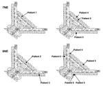
Figure 1. Spot map illustrating case-patient location on the northwestern wing of the eighth floor (8NE) and the seventh floor (7NE) from the 2001 (left panel) and 2003 outbreaks (right panel) at the...
The spot map and time line showed that cases clustered mainly along 2 corridors on 2 floors, 1 directly above the other, around the time of diagnosis. In the 2001 and 2003 outbreaks, all case-patients resided in the same or adjacent rooms before diagnosis (Figure 1).
Environmental air sampling performed 2 months after the last case occurred in 2003 found no A. ustus isolates in the rooms of HSCT patients. One environmental A. ustus isolate was obtained from the carpeted floor of the hall near the room in which the colonized lung transplant recipient resided. The same bronchoscope was used to evaluate each patient; however, it was cleaned after each examination. Also, several patients who were not found to have A. ustus on bronchoalveolar lavage underwent bronchoscopic examination before case-patients, suggesting that cross-contamination was unlikely.
Analyses of Isolates
Figure 2
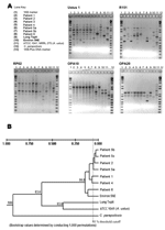
Figure 2. Molecular typing of Aspergillus ustus isolates by using random amplification of polymorphic DNA. The isolate from patient 3 was not viable on subculturing and, as such, was not available for molecular...
Eleven A. ustus isolates were available for analysis. One patient (patient 3) did not have a viable isolate stored, and 1 patient (patient 5) had 3 isolates recovered during the course of infection. A total of 73 bands were resolved from the 11 A. ustus isolates (Figure 2). The isolates recovered from the 5 HSCT were genetically similar. Three isolates from patient 5 were genetically most similar to the isolate from patient 2. At the time of his diagnosis and death, patient 2 resided in a room directly adjacent to and above the room of patient 5, albeit 2 years earlier (Figure 1). Similarly, the isolate from patient 1 was genetically most similar to that of patient 4; patients 1 and 4 resided in adjacent rooms, also separated by a period of 2 years. Of note, the lung transplant patient appeared to be colonized with a strain of A. ustus that was genetically as distant from the patient isolates as the wild-type ATCC strain. Antifungal drug susceptibility testing of clinical isolates demonstrated relatively high MICs to all antifungal drugs tested (Table 2).
We report the first outbreak of disease caused by an unusual fungal pathogen, A. ustus, a mold that has rarely caused invasive disease in humans. This observation is important, given the possibility of common-source acquisition of a potentially antifungal drug–resistant organism.
All members of the A. ustus complex have similar shape with subtle differences. Macroscopically, the colonies appear drab olive to dull brown or gray and woolly with occasional dark purple or yellow exudates. Microscopically, the conidia are large (3.0–4.5 μm) and are rough-walled. Elongate and irregular-shaped Hülle cells that are resistant to desiccation may also be produced (23). A. ustus is toxigenic and produced several mycotoxins such as austdiol, austin, austocystin A, and sterigmatocystin (24–27). Although these toxins may be medically important, the quantities of toxin produced in the environment may not be significant (28,29). The spectrum of disease reported due to A. ustus includes onychomycosis, otitis media, primary cutaneous infection, endocarditis, pneumonia, and disseminated infection, the latter cases occurring largely among immunocompromised hosts such as HSCT recipients. All previously reported cases occurred sporadically in diverse medical centers (Table A1). Many reported cases have been either primary cutaneous disease or disseminated infection, however, we cannot draw firm conclusions regarding the types of infections this organism causes because of the high likelihood of reporting bias. The relative pathogenicity of this Aspergillus species has not been well studied.
In the 6 HSCT patients described in this article, infection developed late after HSCT, with a high proportion of deaths (30,31). These patients also possessed classic risk factors for IA in that most had graft-versus-host disease that required corticosteroid and other immunosuppressive therapy (30). Overall death rates of patients with A. ustus infection was high in this cohort, as in previous cases (4–15). Whether death was attributable to the fungal infection, coinfections, or underlying diseases is unclear.
A common source for the A. ustus infections appears possible, since case-patients clustered in space and time, and a high degree of genetic similarity was noted between isolates from case-patients. Since these patients resided in rooms within close proximity, common source acquisition (e.g., air, water, or surface) is credible. Common source acquisition may not be precluded by case isolate separation in time as Aspergillus conidia are resistant to harsh conditions, surviving in the environment for many years in dormant phase (32). However, the environmental niche of this fungus is not known. Patients were in and out of the hospital after transplantation, so infection could have been acquired in the environment. We also cannot rule out the possibility that other clinical factors (e.g., changes in hosts or antifungal drug administration) selected for specific A. ustus isolates in the patients.
The results of molecular analyses suggest genetic similarity of isolates recovered from patients. Although discriminatory power of RAPD analyses has some limitations (33), the composite analysis demonstrated large separations between patient isolates and the control ATCC strain. Our study is limited by the lack of local environmental A. ustus isolates available for genetic comparison. Although the clinical isolates appear different from the ATCC strain, the genetic similarity of case strains may represent a strain common to our local environment. Also, these analyses are limited by our lack of knowledge concerning A. ustus's modes of reproduction. Specifically, genomic rearrangement with recombination, which has been postulated to occur in several species of Aspergillus, may increase the variation observed between related strains (34).
Our investigation was limited by constraints in conducting retrospective analyses. More timely environmental sampling may have captured more environmental A. ustus isolates (32,35). For example, swabbing of dust-ridden surfaces may have indicated the underlying air quality in terms of fungal spores in the preceding months when infection may have occurred. In the absence of substantial air disturbances, A. ustus spores would be more likely to quickly settle in such areas, given their large size and relatively decreased buoyancy. Construction, a well-known environmental risk factor for IA (36), was ongoing outside the hospital during the time of these outbreaks. We cannot comment on the role of water as a source of infection, which has been reported in multiple hospitals (32,35). Determining the source of infection is further complicated in that a combination of inoculum effect and underlying host immunosuppression make calculating the incubation period problematic. Thus, the source of A. ustus infection among our patients, and whether the infections were nosocomial or community acquired, remains unknown.
These outbreaks of A. ustus infections may be of infection control importance, as the clinical isolates exhibited low susceptibilities to multiple antifungal drugs, as was reported previously (12,17). Although we do not know breakpoints of A. fumigatus resistance, results of prior studies suggest that infection with organisms requiring high MICs of amphotericin or itraconazole is associated with poor clinical outcomes (37,38). Most of our patients received mold-active azole drugs before diagnosis as either prophylaxis or therapy for a previous infection with A. fumigatus. Similarly, A. ustus was recently reported to cause "breakthrough" infection during administration of voriconazole and caspofungin (17). Drug exposure may select for colonization or infection with resistant isolates or facilitate acquired resistance within a colonizing strain. The latter may occur in A. fumigatus isolates exposed to azole antifungal agents (39,40). In this cohort, several patients who received the combination regimen of voriconazole and caspofungin had A. ustus infection resolve; whether this resolution was due to drug synergy in treating relatively resistant organisms is worthy of further consideration.
A. ustus is rare; however, it may be emerging as a cause of systemic disease among immunocompromised hosts in the appropriate setting. A combination of factors, including severity of underlying host immunosuppression and common source acquisition, likely played a role in the reported outbreaks. Active laboratory, environmental, and clinical-based surveillance for A. ustus has been implemented at our hospital based on the results of this investigation; no additional isolates have been identified subsequently. Such intensive monitoring may show similar outbreaks in other facilities. This study also emphasizes the importance of establishing microbial diagnoses to the species level; information obtained is important for infection control and, possibly, to guide antifungal therapies. More studies will be necessary to determine the clinical consequence of antifungal resistance in A. ustus isolates.
Dr. Panackal is an infectious disease fellow at the University of Washington and the Fred Hutchinson Cancer Research Center. His interests include the epidemiology of fungal infections.
Acknowledgments
We thank Estella Whimbey and Nancy Whittington for their help with acquiring information on hospital airflow and construction activity, Robin Olsen for performing the environmental air sampling, Chris Davis for database support, David Madtes and Pat McDowell for obtaining bronchoscopic information, and S. Arunmozhi Balajee and Jennifer Gribskov for their assistance in antifungal drug susceptibility testing.
Financial support was in part provided by NIH grant R21 #AI55928. Dr Panackal received grant support from the 2004 John P. Utz Postdoctoral Fellowship in Medical Mycology sponsored by the National Foundation for Infectious Diseases and Pfizer Inc.
References
- McNeil MM, Nash SL, Hajjeh RA. Trends in mortality due to invasive mycotic diseases in the United States, 1980–1997. Clin Infect Dis. 2001;33:641–7. DOIPubMedGoogle Scholar
- Warnock DA, Hajjeh RA, Lasker BA. Epidemiology and prevention of invasive aspergillosis. Curr Infect Dis Rep. 2001;3:507–16. DOIPubMedGoogle Scholar
- Marr KA, Carter RA, Crippa F, Wald A, Corey L. Epidemiology and outcome of mould infections in hematopoietic stem cell transplant recipients. Clin Infect Dis. 2002;34:909–17. DOIPubMedGoogle Scholar
- Lawrence T, Schockman AT, McVaugh H III. Aspergillus infection of aortic prosthetic valves. Chest. 1971;60:406–14. DOIPubMedGoogle Scholar
- Sandner VK, Schönborn C. Schimmelpilzinfektion der Haut bei ausgedehnter Verbrennung. Deutsch Ges-Wesen. 1973;28:125–8.
- Carrizosa J, Levison ME, Lawrence T, Kaye D. Cure of Aspergillus ustus endocarditis on prosthetic valve. Arch Intern Med. 1974;133:486–90. DOIPubMedGoogle Scholar
- Weiss LM, Thiemke WA. Disseminated Aspergillus ustus infection following cardiac surgery. Am J Clin Pathol. 1983;80:408–11.PubMedGoogle Scholar
- Stiller MJ, Teperman L, Rosenthal SA, Primary cutaneous infection by Aspergillus ustus in a 62-year-old liver transplant recipient. J Am Acad Dermatol. 1994;31:344–7. DOIPubMedGoogle Scholar
- Bretagne S, Marmorat-Khuong A, Kuentz M, Latge JP, Bart-Delabesse E, Cordonnier C. Serum Aspergillus galactomannan antigen testing by sandwich ELISA: practical use in neutropenic patients. J Infect. 1997;35:7–15. DOIPubMedGoogle Scholar
- Ricci RM, Evans JS, Meffert JJ, Kaufman L, Sadkowski LC. Primary cutaneous Aspergillus ustus infection: secondary reported case. J Am Acad Dermatol. 1998;38:797–8. DOIPubMedGoogle Scholar
- Iwen PC, Rupp ME, Bishop MR, Rinaldi MG, Sutton DA, Tarantolo S, Disseminated aspergillosis caused by Aspergillus ustus in a patient following allogeneic peripheral stem cell transplantation. J Clin Microbiol. 1998;36:3713–7.PubMedGoogle Scholar
- Verweij PE, Van den Bergh MFQ, Rath PM, De Pauw BE, Voss A, Meis JFGM. Invasive aspergillosis caused by Aspergillus ustus: case report and review. J Clin Microbiol. 1999;37:1606–9.PubMedGoogle Scholar
- Gené J, Azón-Masoliver A, Guarro J. Cutaneous infection caused by Aspergillus ustus, an emerging opportunistic fungus in immunocompromised patients. J Clin Microbiol. 2001;39:1134–6. DOIPubMedGoogle Scholar
- Nakai K, Kanda Y, Mineishi S. Primary cutaneous aspergillosis caused by Aspergillus ustus following reduced-intensity stem cell transplantation. Ann Hematol. 2002;81:593–6. DOIPubMedGoogle Scholar
- Azzola P Jr, Habicht JM. Use of lung resection and voriconazole for successful treatment of invasive pulmonary Aspergillus ustus infection. J Clin Microbiol. 2004;42:4805–8. DOIPubMedGoogle Scholar
- Baddley JW, Stroud TP, Salzman D, Pappas PG. Invasive mold infections in allogeneic bone marrow transplant recipients. Clin Infect Dis. 2001;32:1319–24. DOIPubMedGoogle Scholar
- Pavie J, Lacroix C, Hermose DG. Breakthrough disseminated Aspergillus ustus infection in allogeneic hematopoietic stem cell transplant recipients receiving voriconazole and caspofungin prophylaxis. J Clin Microbiol. 2005;43:4902–4. DOIPubMedGoogle Scholar
- Ascioglu S, Rex JH, de Pauw J. Defining opportunistic invasive fungal infections in immunocompromised patients with cancer and hematopoietic stem cell transplants: an international consensus. Clin Infect Dis. 2002;34:7–14. DOIPubMedGoogle Scholar
- Rath PM, Petermeier K, Verweij PE, Ansorg R. Differentiation of Aspergillus ustus strains by random amplification of polymorphic DNA. J Clin Microbiol. 2002;40:2231–3. DOIPubMedGoogle Scholar
- Sneath PHA, Sokal RR, eds. Numerical taxonomy: the principles and practice of numerical classification. San Francisco: W.H. Freeman; 1973.
- Pfaller MA, Chaturvedi V, Espinel-Ingroff A. Reference method for broth dilution antifungal susceptibility testing of yeasts; approved standard. National Committee for Clinical Laboratory Standards document M27-A, 22:1-30. The Committee; 2003.
- Bartizal C, Odds FC. Influences of methodological variables on susceptibility testing of caspofungin against Candida species and Aspergillus fumigatus. Antimicrob Agents Chemother. 2003;47:2100–7. DOIPubMedGoogle Scholar
- Sutton DA, Fothergill AW, Rinaldi MG. Aspergillus ustus. In: Guide to clinically significant fungi. 1st ed. Baltimore: Lippincott Williams and Wilkins; 1998.
- Kfir R, Johannsen E, Vleggaar R. Mutagenic activity of austocystins—secondary metabolites of Aspergillus ustus. Bull Environ Contam Toxicol. 1986;37:643–50. DOIPubMedGoogle Scholar
- Rabie CJ, Steyn M, van Schalkwyk GC. New species of Aspergillus producing sterigmatocystin. Appl Environ Microbiol. 1977;33:1023–5.PubMedGoogle Scholar
- Chexal KK, Spinger JP, Clardy J. Austin, a novel polyisoprenoid mycotoxin from Aspergillus ustus. J Am Chem Soc. 1976;98:6748. DOIPubMedGoogle Scholar
- Vleggaar R, Steyn PS, Nagel DW. Constitution and absolute configuration of austdiol, the main toxic metabolite from Aspergillus ustus. J Chem Soc [Perkin 1]. 1974;1:45–9.
- Nielsen KF, Gravesen S, Nielsen PA. Production of mycotoxins on artificially and naturally infested building materials. Mycopathologia. 1999;145:43–56. DOIPubMedGoogle Scholar
- Nielsen KF. Mycotoxin production by indoor molds. Fungal Genet Biol. 2003;39:103–17. DOIPubMedGoogle Scholar
- Marr KA, Carter RA, Boeckh M, Martin P, Corey L. Invasive aspergillosis in allogeneic stem cell transplant recipients: changes in epidemiology and risk factors. Blood. 2002;100:4358–66. DOIPubMedGoogle Scholar
- Fukuda T, Boeckh M, Carter RA. Risks and outcomes of invasive fungal infections in recipients of allogeneic hematopoietic stem cell transplants after nonmyeloablative conditioning. Blood. 2003;102:827–33. DOIPubMedGoogle Scholar
- Warris A, Klaassen CHW, Meis JFGM. Molecular epidemiology of Aspergillus fumigatus isolates recovered from water, air, and patients shows two clusters of genetically distinct strains. J Clin Microbiol. 2003;41:4101–6. DOIPubMedGoogle Scholar
- Lasker BA. Evaluation of performance of four genotypic methods for studying the genetic epidemiology of Aspergillus fumigatus isolates. J Clin Microbiol. 2002;40:2886–92. DOIPubMedGoogle Scholar
- Geiser DM, Timberlake WE, Arnold ML. Loss of meiosis in Aspergillus. Mol Biol Evol. 1996;13:809–17.PubMedGoogle Scholar
- Anaissie EJ, Stratton SL, Dignani MC. Pathogenic Aspergillus species recovered from a hospital water system: a 3-year prospective study. Clin Infect Dis. 2002;34:780–9. DOIPubMedGoogle Scholar
- Hajjeh RA, Warnock DW. Counterpoint: invasive aspergillosis and the environment—rethinking our approach to prevention. Clin Infect Dis. 2001;33:1549–52. DOIPubMedGoogle Scholar
- Lass-Florl. Kofler G, Kropshofer G. In-vitro testing of susceptibility to amphotericin B is a reliable predictor of clinical outcome in invasive aspergillosis. J Antimicrob Chemother. 1998;42:497–502. DOIPubMedGoogle Scholar
- Denning DW, Radford SA, Oakley KL, Hall L, Johnson EM, Warnock DW. Correlation between in-vitro susceptibility testing to itraconazole and in-vivo outcome of Aspergillus fumigatus infection. J Antimicrob Chemother. 1997;40:401–14. DOIPubMedGoogle Scholar
- Dannaoui E, Borel E, Monier MF, Piens MA, Picot S, Persat F. Acquired itraconazole resistance in Aspergillus fumigatus. J Antimicrob Chemother. 2001;47:333–40. DOIPubMedGoogle Scholar
- Warris A, Weemaes CM, Verweij PE. Multidrug resistance in Aspergillus fumigatus. N Engl J Med. 2002;347:2173–4. DOIPubMedGoogle Scholar
Figures
Tables
Cite This ArticleTable of Contents – Volume 12, Number 3—March 2006
| EID Search Options |
|---|
|
|
|
|
|
|
Please use the form below to submit correspondence to the authors or contact them at the following address:
Kieren A. Marr, Fred Hutchinson Cancer Research Center, University of Washington, Seattle, 1100 Fairview Ave D3-100, Seattle, WA 98109, USA; fax: 206-667-4411
Top