Volume 12, Number 8—August 2006
Dispatch
Human Metapneumovirus, Australia, 2001–2004
Cite This Article
Citation for Media
Abstract
We examined 10,025 respiratory samples collected for 4 years (2001–2004) and found a 7.1% average annual incidence of human metapneumovirus. The epidemic peak of infection was late winter to spring, and genotyping showed a change in predominant viral genotype in 3 of the 4 years.
Human metapneumovirus (HMPV) is now recognized as a substantial cause of acute respiratory infection, particularly in children; it has phenotypic and clinical characteristics that are similar to those of respiratory syncytial virus (RSV) (1,2). HMPV is ubiquitous; it infects most children at an early age, and distinct epidemic peaks are reported in the winter months (1,3–6). However, many of theses studies were conducted in the Northern Hemisphere and involved samples collected during a relatively short period; few data exist from extended studies over several years that involve populations in other parts of the world.
Although 2 main HMPV types are recognized (A and B) (1,3,7), each with 2 subtypes (A1, A2; B1, B2) (8), the extent of genetic variation of circulating HMPV subtypes over time has not been extensively examined. We sought to determine the incidence and seasonal distribution of HMPV in an Australian population from 2001 to 2004 and to establish the pattern of genotype distribution during those 4 years by examining the genetic variability of the P gene in 640 of 707 HMPV-positive samples.
The necessary ethics approval for this study was obtained from the Royal Children's Hospital ethics committee. We collected nasopharyngeal aspirate (NPA) specimens from January 2001 through December 2004 from patients with acute lower respiratory tract infection in Queensland, Australia. Patients were from 3 months to 93 years of age (mean 8.2 years, median 1.37 years), and 78.1% of specimens were from children <5 years of age. Nucleic acids were extracted from 0.2 mL of each NPA specimen by using the High Pure Viral Nucleic Acid kit (Roche Diagnostics, Mannheim, Germany), according to the manufacturer's instructions. Extracts were analyzed for HMPV sequences by reverse transcriptase PCR (9). For samples collected during 2001 and 2002, other viral respiratory pathogens were detected by using a direct fluorescent antibody assay (DFA) in combination with a culture-augmented DFA method (10). For samples collected in 2003 and 2004, these pathogens were detected by multiplex PCR (10).
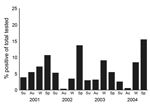
Of 10,025 NPA specimens tested, 707 were positive for HMPV, for an overall incidence of 7.1% during the 4 years. The youngest HMPV-positive patient was 4 months old, and the oldest was 79 years. In children (<18 years of age) the incidence of virus was 7.4%, and 91.9% of HMPV-positive children were <5 years of age. The seasonal distribution of HMPV infection showed a distinctive pattern for each of the 4 years studied (Figure 1). In 2001, HMPV showed broad seasonal activity; incidence was >5% in 3 consecutive seasons (autumn, winter, and spring) and peaked at 10.6% in the spring (September–November). In 2002 and 2004, most HMPV activity was in spring, (incidence of 13.6% and 15.4%, respectively), with little evidence during autumn (March–May). In 2003, the peak incidence of 9.0% occurred in winter (June–August) and persisted into spring (5.4%). In all years, virus was present well into summer (December–February), with an incidence ranging from 2.5% to 5.2%. On examination of those samples collected in 2003 and 2004, which were all previously analyzed for common respiratory viruses by PCR, HMPV was the most frequently detected respiratory virus in children during the spring of each year. Expressed as an annual average over the 4 years studied, the predominant viral pathogen was RSV (9.2%), followed by HMPV (7.1%), influenza A (3.5%), parainfluenza virus 3 (2.3%), and adenovirus (1.3%). In 6.8% of HMPV-positive cases, evidence of co-infection with another respiratory virus was seen; 20 patients were concurrently infected with an adenovirus, 10 with influenza A virus, 8 with RSV, 9 with parainfluenza virus 3, and 1 with parainfluenza virus 2.
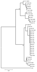
Amplification products generated directly from 640 HMPV-positive NPA specimens were genotyped as previously described (11) (GenBank accession nos. DQ112292–DQ112320 and DQ121378–DQ121384). Data showed that all 4 viral subtypes cocirculated during each of the 4 years studied (Table 1, Figure 2). However, a different subtype predominated during 3 of the 4 years: HMPV subtype A1 was dominant in 2001, subtype A2 in 2002 and 2003, and subtype B1 in 2004 (Table 1).
Clinical records from 273 patients who were positive for HMPV were scrutinized, and data describing clinical features and length of hospital stay were recorded. Of these patients, 203 (74.4%) were admitted to hospital with a median length of admission of 3 days and a mean of 6.5 days. The predominant clinical features were cough (63%), rhinorrhea (61%), respiratory crackles/crepitations (60%), and fever (57%) (Table 2). Ninety (33.9%) of the 273 patients had a chest radiograph, and 77 (85.6%) patients showed bilateral parahilar peribronchial infiltrates consistent with a lower respiratory tract infection. Disease severity of the 273 HMPV-positive patients was classified as mild (46.8%), moderate (42.5%), and severe (10.7%), based on the use of supplemental oxygen and fluids and length of hospital stay.
This study of HMPV infection is the largest so far reported. The results of recent, similar studies suggested that peak periods of infection with HMPV predominate during winter in the Northern Hemisphere. However, this finding has not been extensively examined over an extended period with a large, continuous sample. Our study found that the peak period of HMPV infection in Queensland, Australia, occurs predominantly in spring (August–October) but that HMPV can be detected in every month. This finding suggests that HMPV activity, like RSV activity, occurs in the community throughout the year, and peaks of infection are a result of seasonal environmental factors.
Although RSV predominated in all years, HMPV was the second most frequently detected virus in each year studied. The low rate of co-infection of HMPV with other respiratory viruses (including RSV) suggests that co-infection may not be common in our community. When analyzing disease severity in this sample with a 2-sided test of proportions, we saw no significant difference between patients with a co-infection and those without. Therefore, our data did not support the suggestion by others that co-infection of HMPV with RSV or other viral respiratory pathogens is a risk factor for severe disease (6).
The shift in predominant HMPV genotype observed in this study was similar to those reported for RSV and influenza viruses (6) and can be attributed to changes in immunity of the population in response to antigenic differences between the predominant circulating strains (12,13). However, a relationship between genotype and disease severity, as previously established for RSV (14,15), did not appear to apply for HMPV, but we plan to examine this relationship further.
The clinical features associated with HMPV infection in this study were not sufficiently distinctive to clinically differentiate it from other respiratory viral infections in children, particularly those attributed to RSV. In addition, few patients (10.6%) had severe disease, but most (76%) were sufficiently ill to be admitted and treated in the hospital for >3 days, which represents a substantial amount in healthcare costs.
Finally, this comprehensive study, conducted for 48 months, is the first one aimed at establishing an accurate estimate of the incidence and seasonal distribution of HMPV infection and to determine the genetic variation of HMPV circulating in our population. The clinical spectrum of infection in a substantial proportion of HMPV-positive patients has been described, and studies are continuing to fully elucidate the clinical effect of infection with this virus in our community.
Dr Sloots is the director of the research section of the Queensland Paediatric Infectious Diseases Laboratory at Sir Albert Sakzewski Virus Research Centre. His main research interests are in pediatric infectious diseases, particularly viral respiratory disease.
Acknowledgments
We thank the staff of the Microbiology Division of the Queensland Health Pathology Service, Royal Brisbane and Women's Hospital. We also acknowledge the Directors of Paediatrics, Queensland Health, who gave permission to review the medical records of HMPV-positive patients.
This study was supported by the Royal Children's Hospital Foundation grants I 922-034 and R 912-009, the Woolworths Fresh Futures Appeal, and the National Health and Medical Research Council, Australia, project grant 243702.
References
- van den Hoogen BG, de Jong JC, Groen J, Kuiken T, de Groot R, Fouchier RA, A newly discovered human pneumovirus isolated from young children with respiratory tract disease. Nat Med. 2001;7:719–24. DOIPubMedGoogle Scholar
- Nissen MD, Siebert DJ, Mackay IM, Sloots TP, Withers SJ. Evidence of human metapneumovirus in Australian children. Med J Aust. 2002;176:188.PubMedGoogle Scholar
- Bastien N, Ward D, van Caeseele P, Brandt K, Lee SH, McNabb G, Human metapneumovirus infection in the Canadian population. J Clin Microbiol. 2003;41:4642–6. DOIPubMedGoogle Scholar
- Freymouth F, Vabret A, Legrand L, Eterradossi N, Lafay-Delaire F, Brouard J, Presence of the new human metapneumovirus in French children with bronchiolitis. Pediatr Infect Dis J. 2003;22:92–4. DOIPubMedGoogle Scholar
- Peiris JS, Tang WH, Chan KH, Khong PL, Guan Y, Lau YL, Children with respiratory disease associated with metapneumovirus in Hong Kong. Emerg Infect Dis. 2003;9:628–33.PubMedGoogle Scholar
- Robinson JL, Lee BE, Bastien N, Li Y. Seasonality and clinical features of human metapneumovirus infection in children in northern Alberta. J Med Virol. 2005;76:98–105. DOIPubMedGoogle Scholar
- van den Hoogen BG, Bestebroer TM, Osterhaus AD, Fouchier RA. Medline Analysis of the genomic sequence of a human metapneumovirus. Virology. 2002;295:119–32. DOIPubMedGoogle Scholar
- Boivin G, Mackay IM, Sloots TP, Madhi S, Freymuth F, Wolf D, Global genetic diversity of human metapneumovirus fusion gene. Emerg Infect Dis. 2004;10:1154–7.PubMedGoogle Scholar
- Mackay IM, Bialasiewicz S, Jacob KC, McQueen E, Arden KA, Nissen MD, Genetic diversity of human metapneumovirus over 4 consecutive years in Australia. J Infect Dis. 2006;193:160–3. DOIPubMedGoogle Scholar
- Syrmis MW, Whiley DM, Thomas M, Mackay IM, Williamson J, Siebert DJ, A sensitive, specific, and cost-effective multiplex reverse transcriptase-PCR assay for the detection of seven common respiratory viruses in respiratory samples. J Mol Diagn. 2004;6:125–31. DOIPubMedGoogle Scholar
- Mackay IM, Bialasiewicz S, Waliuzzaman Z, Chidlow GR, Fegredo DC, Laingam S, Use of the P gene to genotype human metapneumovirus identifies four viral subtypes. J Infect Dis. 2004;190:1913–8. DOIPubMedGoogle Scholar
- Peret TC, Hall CB, Schnabel KC, Golub JA, Anderson LJ. Circulation patterns of genetically distinct group A and B strains of human respiratory syncytial virus in a community. J Gen Virol. 1998;79:2221–9.PubMedGoogle Scholar
- Cane PA, Pringle CR. Evolution of subgroup A respiratory syncytial virus: evidence for progressive accumulation of amino acid changes in the attachment protein. J Virol. 1995;69:2918–25.PubMedGoogle Scholar
- Kneyber MC, Brandenburg AH, Rothbarth PH, de Groot R, Ott A, van Steensel-Moll HA. Relationship between clinical severity of respiratory syncytial virus infection and subtype. Arch Dis Child. 1996;75:137–40. DOIPubMedGoogle Scholar
- Walsh EE, McConnochie KM, Long CE, Hall CB. Severity of respiratory syncytial virus infection is related to virus strain. J Infect Dis. 1997;175:814–20. DOIPubMedGoogle Scholar
Figures
Tables
Cite This Article1These authors contributed equally to this study.
Table of Contents – Volume 12, Number 8—August 2006
| EID Search Options |
|---|
|
|
|
|
|
|


Please use the form below to submit correspondence to the authors or contact them at the following address:
Theo P. Sloots, Queensland Pediatric Infectious Diseases Laboratory, Sir Albert Sakzewski Virus Research Centre, Royal Children’s Hospital and Health Service District, Herston Rd, Herston, Queensland, Australia 4029
Top