Volume 13, Number 12—December 2007
Research
Susceptibility of Canada Geese (Branta canadensis) to Highly Pathogenic Avian Influenza Virus (H5N1)
John Pasick* , Yohannes Berhane*, Carissa Embury-Hyatt*, John Copps*, Helen Kehler*, Katherine Handel*, Shawn Babiuk*, Kathleen Hooper-McGrevy*, Yan Li†, Quynh Mai Le‡, and Song Lien Phuong§
Figure 2
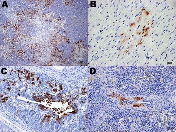
Figure 2. Immunohistochemical (IHC) staining for influenza virus nucleoprotein in tissues of naïve juvenile Canada geese after challenge with influenza virus (H5N1). A) Pancreas. Large areas of necrosis are surrounded by pancreatic acinar cells with strong positive intranuclear and intracytoplasmic immunolabeling. B) Heart. Positive intranuclear and intracytoplasmic immunolabeling of myocytes. C) Proventriculus. Strong positive immunolabeling of compound tubular gland epithelium. D) Splenic arteriole. Positive IHC staining of vascular smooth muscle cells.
Page created: July 06, 2010
Page updated: July 06, 2010
Page reviewed: July 06, 2010
The conclusions, findings, and opinions expressed by authors contributing to this journal do not necessarily reflect the official position of the U.S. Department of Health and Human Services, the Public Health Service, the Centers for Disease Control and Prevention, or the authors' affiliated institutions. Use of trade names is for identification only and does not imply endorsement by any of the groups named above.