Volume 13, Number 2—February 2007
Letter
Frog Virus 3 Infection, Cultured American Bullfrogs
Debra L. Miller* , Sreekumari Rajeev*, Matthew J. Gray†, and Charles A. Baldwin*
Figure
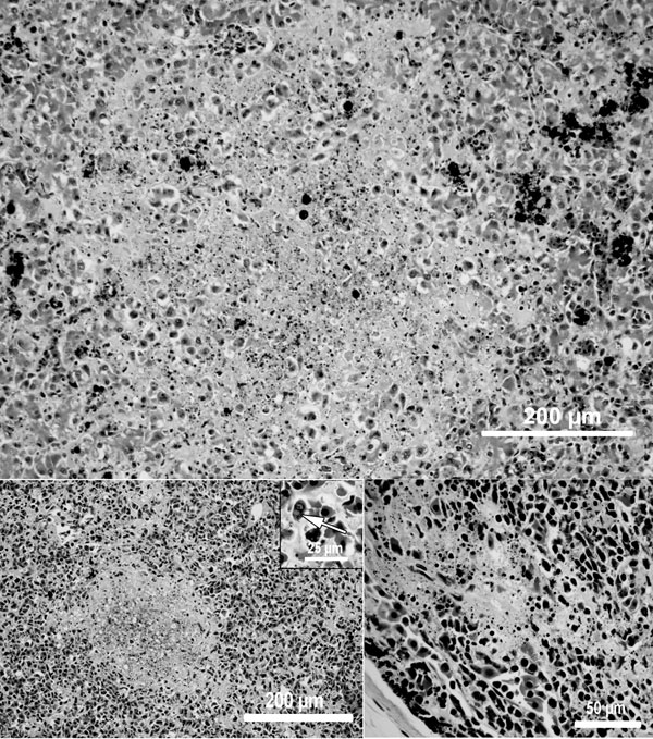
Figure. Light microscopic images of the liver (A), spleen (B), and thymus (C) showing necrosis in an American bullfrog (Rana catesbeiana) metamorph infected with frog virus 3. Spleen (B) inset shows intracytoplasmic viral inclusion bodies. Hematoxylin and eosin stain.
Page created: June 29, 2010
Page updated: June 29, 2010
Page reviewed: June 29, 2010
The conclusions, findings, and opinions expressed by authors contributing to this journal do not necessarily reflect the official position of the U.S. Department of Health and Human Services, the Public Health Service, the Centers for Disease Control and Prevention, or the authors' affiliated institutions. Use of trade names is for identification only and does not imply endorsement by any of the groups named above.