Volume 14, Number 11—November 2008
Dispatch
Pyemotes ventricosus Dermatitis, Southeastern France
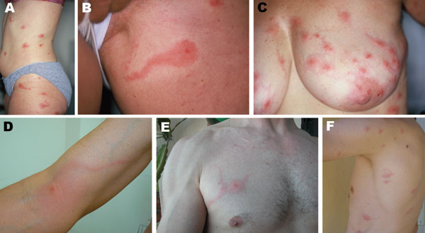
Figure 1

Figure 1. A–F) Photographs of 6 persons with skin lesions of Pyemotes ventricosus dermatitis. Note the central microvesicles, ulcerations or crusts, and some lesions with the comet sign. D) Lymphangitis-like dermatitis. E, F) Lesions resulting from natural infection of 2 of the investigators.
Page created: July 18, 2010
Page updated: July 18, 2010
Page reviewed: July 18, 2010
The conclusions, findings, and opinions expressed by authors contributing to this journal do not necessarily reflect the official position of the U.S. Department of Health and Human Services, the Public Health Service, the Centers for Disease Control and Prevention, or the authors' affiliated institutions. Use of trade names is for identification only and does not imply endorsement by any of the groups named above.