Volume 14, Number 12—December 2008
Letter
Parachlamydia acanthamoebae Infection and Abortion in Small Ruminants
Figure

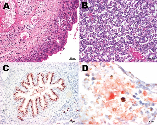
Figure. A) Sheep placenta positive by real-time PCR and immunohistochemistry for Parachlamydia spp. and Chlamydiaceae. Chlamydophila abortus was identified by ArrayTube Microarray. Necrotizing placentitis and vasculitis are shown (hematoxylin and eosin stain; magnification ×200). B) Fetal lung of the sheep abortion specimen positive by real-time PCR and immunohistochemical tests for Parachlamydia spp. and Chlamydiaceae; interstitial pneumonia is shown (hematoxylin and eosin stain; magnification ×200). C) Fetal lung that was positive by real-time PCR and immunohistochemical testing for Parachlamydia spp. Positive granular material can be seen within the lung tissue. Antigen detection (immunohistochemistry) was carried out with a polyclonal antibody directed against Parachlamydia spp. 3-amino-9-ethylcarbazole/peroxidase method (hematoxylin counterstain; magnification ×200). D) Double immunohistochemical labeling of the sheep placenta that was positive by real-time PCR and immunohistochemical tests for Chlamydiaceae and Parachlamydia spp. The simultaneous presence of Chlamydiaceae and Parachlamydia spp. granular reaction is shown within necrotic trophoblastic epithelium and neutrophilic exudate (diaminobenzidine/AEC/peroxidase method, hematoxylin counterstain; magnification ×1,000).