Volume 14, Number 12—December 2008
Letter
Candidate New Species of Kobuvirus in Porcine Hosts
Cite This Article
Citation for Media
To the Editor: Picornaviruses (family Picornaviridae) are small, nonenveloped viruses with single-stranded, positive-sense genomic RNA, currently divided into 8 genera: Enterovirus, Aphthovirus, Cardiovirus, Hepatovirus, Parechovirus, Erbovirus, Teschovirus, and Kobuvirus (1). To date, the genus Kobuvirus consists of 2 species, Aichi virus and Bovine kobuvirus, each possessing 1 serotype. Aichi virus (strain A846/88) was first isolated from a stool sample obtained from a person with acute gastroenteritis in 1991 (2). Bovine kobuvirus (strain U-1) was detected in bovine sera and in feces samples from clinically healthy cattle in 2003 (3). Human and bovine kobuviruses were first isolated in Japan. Recently, kobuviruses have also been detected in humans in other countries in Asia (4), Europe (5,6), and South America (5) and in calves with diarrhea in Thailand (7). The Aichi virus and bovine kobuvirus genomes are approximately 8.2–8.3 kb, respectively, and both have a typical picornavirus genome organization, including the L protein following structural (VP0, VP3, and VP1) and nonstructural (2A–2C and 3A–3D) regions (1,3). Genetic identity between Aichi and U-1 viruses ranges from 47.7% (3′ untranslated region) through 70.8% (3D region) (3). In this study, we report a new candidate species of kobuvirus. Porcine kobuvirus was serendipitously detected in fecal samples from domestic pigs in Hungary.
Fecal samples were collected in February 2007 from 15 healthy piglets (Sus scrofa domestica) <10 days of age from a farm in Ebes located in eastern Hungary. The aim of the study was to detect porcine calicivirus (norovirus and sapovirus) in domestic pigs by using reverse transcription–PCR (RT-PCR), using the generic primer pairs p289/p290 designed for the calicivirus RNA-dependent RNA polymerase gene (319 nt for norovirus or 331 nt for sapovirus) (8). RNA isolation and RT-PCR were performed as described previously (9). PCR products were sequenced directly in both directions with the BigDye Terminator Cycle Sequencing Ready Reaction Kit (PE Applied Biosystems, Warrington, UK) by using the PCR primers and run on an automated sequencer (ABI PRISM 310 Genetic Analyzer; Applied Biosystems, Stafford, TX, USA). Phylogenetic analysis was conducted by using MEGA software version 4.0 (10). Complete nucleotide sequence of porcine kobuvirus (strain Kobuvirus/swine/S-1-HUN/2007/Hungary) was submitted to GenBank under accession no. EU787450.
Figure
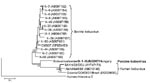
Figure. Phylogenetic tree of porcine kobuvirus (Kobuvirus/swine/S-1-HUN/2007/Hungary, GenBank accession no. EU787450), based upon the 1,065-nt fragment of the kobuvirus 3C/3D regions. The phylogenetic tree was constructed by using the neighbor-joining clustering method;...
Two (13.3%) of 15 samples were positive for porcine sapoviruses; however, a consequent nonspecific, ≈1,100-nt, strong, and single PCR product was found in all samples by agarose gel electrophoresis (data not shown). The nucleotide sequence of the 1,065-nt nonspecific PCR product was determined. Genetic and amino acid similarity was found to be bovine (U-1) and human Aichi kobuvirus 3C (87 nt) and 3D (978 nt) regions in GenBank database by using BLAST (http://blast.ncbi.nlm.nih.gov/Blast.cgi). Nucleotide and amino acid identity of the 3C–3D regions were 73%–79% and 69%–70% to U-1 strain and Aichi virus, respectively. The phylogenetic tree confirmed that S-1-HUN belonged to kobuviruses and formed a distinct lineage (Figure). Cleavage sites for 3C and 3D was Q/S. Highly conserved amino acid motif KDELR in 3D (RNA-dependent RNA polymerase) region and high rate of cytidine (29%) and uracil (26%) nucleoside composition were seen in the 3C and 3D parts of the genome of strain S-1-HUN; both are suspected to be a typical skew of kobuviruses (3).
Most picornavirus genera consist of >2 species (1). Our study reports detection of kobuvirus in domestic pigs. Serendipitously, the generic calicivirus primers p289 and p290, designed for a calicivirus RNA-dependent RNA polymerase region, amplified a kobuvirus 3C/3D region when specimens were tested for porcine caliciviruses by RT-PCR. Comparison of the primers p289 and p290 and the S-1-HUN sequence showed that there are 12- and 11-bp homologous regions between the kobuvirus and the 3′ end of the primer sequences, respectively. Reverse primer p289 designed for calicivirus (norovirus and sapovirus) conserved amino acid 3D motif YGDD, which is also present in kobuviruses.
All apparently healthy animals <10 days of age carried the kobuvirus, which was excreted in the feces. These results indicate a general circulation and endemic infection of kobuvirus on the tested farm. In addition, because of its analogy to other picornaviruses and because bovine kobuvirus was first detected in culture medium that originated from cattle sera (1,3), we cannot exclude the possibility that the S-1-HUN–like kobuvirus can cause viremia (and generalized infection) in swine. S-1-HUN–like virus may typically cause asymptomatic infections in swine. However, epidemiologic and molecular studies are required regarding the importance of this virus as a causative agent of some diseases of domestic pigs and related animals. Sequence analysis of the complete nucleotide and amino acid sequences of coding (L, P1, P2, and P3: 7,467 nt) and noncoding regions and the genetic organization strain indicate that S-1-HUN is a typical kobuvirus. Phylogenetic analysis shows that S-1-HUN strain is genetically included in the genus Kobuvirus but is distinct from Aichi and bovine kobuviruses. Porcine kobuvirus strain S-1-HUN is a candidate for a new, third species of the genus Kobuvirus.
Acknowledgment
This work was supported by grants from the Hungarian Scientific Research Fund (OTKA, F048433), and the Enteric Virus Emergence, New Tools (EVENT, SP22-CT-2004-502571) funded by the European Union.
References
- Online ICTV. Discussion forum of the International Committee on Taxonomy of Viruses, ICTV 2008 Official Taxonomy [cited 2008 Oct 22]. Available from http://talk.ictvonline.org
- Yamashita T, Kobayashi S, Sakae K, Nakata S, Chiba S, Ishihara Y, Isolation of cytopathic small round viruses with BS-C-1 cells from patients with gastroenteritis. J Infect Dis. 1991;164:954–7.PubMedGoogle Scholar
- Yamashita T, Ito M, Kabashima Y, Tsuzuki H, Fujiura A, Sakae K. Isolation and characterization of a new species of kobuvirus associated with cattle. J Gen Virol. 2003;84:3069–77. DOIPubMedGoogle Scholar
- Pham NT, Khamrin P, Nguyen TA, Kanti DS, Phan TG, Okitsu S, Isolation and molecular characterization of Aichi viruses from fecal specimens collected in Japan, Bangladesh, Thailand, and Vietnam. J Clin Microbiol. 2007;45:2287–8. DOIPubMedGoogle Scholar
- Oh DY, Silva PA, Hauroeder B, Deidrich S, Cardoso DD, Schreier E. Molecular characterization of the first Aichi viruses isolated in Europe and in South America. Arch Virol. 2006;151:1199–206. DOIPubMedGoogle Scholar
- Ambert-Balay K, Lorrot M, Bon F, Giraudon H, Kaplon J, Wolfer M, Prevalence and genetic diversity of Aichi virus strains in stool samples from community and hospitalized patients. J Clin Microbiol. 2008;46:1252–8. DOIPubMedGoogle Scholar
- Khamrin P, Maneekarn N, Peerakome S, Okitsu S, Mizuguchi M, Ushijama H. Bovine kobuviruses from cattle with diarrhea. Emerg Infect Dis. 2008;14:985–6. DOIPubMedGoogle Scholar
- Jiang X, Huang PW, Zhong WM, Farkas T, Cubitt DW, Matson DO. Design and evaluation of a primer pair that detects both Norwalk- and Sapporo-like caliciviruses by RT-PCR. J Virol Methods. 1999;83:145–54. DOIPubMedGoogle Scholar
- Reuter G, Krisztalovics K, Vennema H, Koopmans M. Szűcs Gy. Evidence of the etiological predominance of norovirus in gastroenteritis outbreaks—emerging new variant and recombinant noroviruses in Hungary. J Med Virol. 2005;76:598–607. DOIPubMedGoogle Scholar
- Tamura K, Dudley J, Nei M, Kumar S. MEGA4: molecular evolutionary genetics analysis (MEGA) software version 4.0. Mol Biol Evol. 2007;24:1596–9. DOIPubMedGoogle Scholar
Figure
Cite This ArticleRelated Links
Table of Contents – Volume 14, Number 12—December 2008
| EID Search Options |
|---|
|
|
|
|
|
|
Please use the form below to submit correspondence to the authors or contact them at the following address:
Gábor Reuter, Regional Laboratory of Virology, National Reference Laboratory of Gastroenteric Viruses, ÁNTSZ Regional Institute of State, Public Health Service, Szabadság út 7, H-7623 Pécs, Hungary
Top