Volume 14, Number 7—July 2008
Research
Toxinotype V Clostridium difficile in Humans and Food Animals
Cite This Article
Citation for Media
Abstract
Clostridium difficile is a recognized pathogen in neonatal pigs and may contribute to enteritis in calves. Toxinotype V strains have been rare causes of human C. difficile–associated disease (CDAD). We examined toxinotype V in human disease, the genetic relationship of animal and human toxinotype V strains, and in vitro toxin production of these strains. From 2001 through 2006, 8 (1.3%) of 620 patient isolates were identified as toxinotype V; before 2001, 7 (<0.02%) of ≈6,000 isolates were identified as toxinotype V. Six (46.2%) of 13 case-patients for whom information was available had community-associated CDAD. Molecular characterization showed a high degree of similarity between human and animal toxinotype V isolates; all contained a 39-bp tcdC deletion and most produced binary toxin. Further study is needed to understand the epidemiology of CDAD caused by toxinotype V C. difficile, including the potential of foodborne transmission to humans.
Recent evidence suggests that the epidemiology of Clostridium difficile–associated disease (CDAD) is increasing in incidence and severity (1–3). These changes are due, at least in part, to the emergence of a more virulent C. difficile strain, designated NAP1 (based on its pulsed-field gel electrophoresis [PFGE] pattern), BI (by restriction endonuclease analysis [REA]), toxinotype III (by PCR characterization of the pathogenicity locus), and 027 (by PCR ribotyping) (4). However, the emergence of BI/NAP1/027 may not be solely responsible for changes in CDAD epidemiology, and the origin of this and other virulent strains is still largely unknown. C. difficile has also recently emerged as a pathogen or commensal in food animals such as neonatal pigs and beef and dairy calves (5–7); most of these animal isolates are toxigenic. Although several ribotypes have been identified in calves, the predominant ribotype in both calves and pigs is a toxinotype V strain (8,9). Moreover, recent reports suggest that C. difficile strains recognized as causes of human disease may contaminate retail meats (10).
To better understand whether food animals could be a source of infection for humans, we investigated recent and past human CDAD caused by toxinotype V C. difficile and compared isolates from these cases with those recovered from neonatal pigs and calves. We documented apparent changes in the frequency with which these toxinotype V strains cause human CDAD and compared the molecular characterization and toxin production of these strains with those of recent epidemic (i.e., BI/NAP1/027) and nonepidemic isolates.
Human Case Finding and Definitions
Case finding was performed by reviewing recent and past human isolates of interest from 2 sources. Cases were defined as patients with clinical isolates identified as toxinotype V by analysis of restriction fragment length polymorphisms (RFLPs) of toxin-encoding genes. First, we reviewed 620 C. difficile human isolates sent to the Centers for Disease Control and Prevention (CDC) from healthcare facilities and health departments in multiple states during hospital-associated outbreaks reported from 2001 through early 2007. Second, we reviewed a database of >6,000 isolates maintained by the Hines Veterans Affairs (VA) Hospital, representing CDAD reported from multiple healthcare facilities from 1984 up to January 1, 2001. All toxinotype V isolates were obtained from patients with a diagnosis of CDAD based on clinical history (e.g., diarrhea) and a positive clinical laboratory test for C. difficile toxin (e.g., cytotoxin assay or enzyme immunoassay).
Isolates identified from the Hines VA Hospital database were designated “past,” and isolates sent from hospitals to CDC from 2001 through early 2007 were designated “recent.” Additional clinical information was obtained for recent case-patients from a standard reporting form completed by the original submitting institutions. The proportions of past and recent isolates identified as toxinotype V were compared with the χ2 test results by using SPSS version 13.0 (SPSS, Inc., Chicago, IL, USA).
Case-patients were categorized with regard to likely place of acquisition according to recommendations developed by the Clostridium difficile Surveillance Working Group (11). Cases of CDAD were considered community-associated (CA-CDAD) if symptom onset (or positive C. difficile toxin test) occurred within <48 h after the patient was admitted to a healthcare facility, provided that it had been >12 weeks since the patient was last discharged from a healthcare facility. Cases of CDAD were considered healthcare facility–associated CDAD (HCFA-CDAD) if the patient had symptom onset >48 h after admission to a healthcare facility or was discharged from a healthcare facility within the previous 4 weeks. Cases were classified as indeterminate if symptom onset was within <48 h of a patient’s admission to a healthcare facility and 4–12 weeks since discharge from a previous admission. Case-patients were considered exposed to antimicrobial agents if the patient received a dose of any antimicrobial agent within the 30 days before symptom onset.
Laboratory Methods
C. difficile isolates from humans and animals with clinical disease were obtained from diagnostic laboratories as previously described (4,8). Swine isolates were obtained from neonatal pigs with CDAD in North Carolina, Iowa, Texas, Utah, Ohio, and Arizona from 1999 to 2005. Bovine strains were isolated from January 1, 2003, through 2005, mainly from diarrheic Holstein calves 1 day to 6 weeks of age, originating in southern California, Arizona, New Mexico, Nevada, Texas, and Utah and maintained in pre-feedlot housing in calf ranches in Arizona. Domestic animal isolates were selected for study on the basis of host of origin, date of isolation, and geographic origin, all of which were independent. All food animal and human isolates were typed by PFGE with SmaI digestion as previously described (12,13) and analyzed with BioNumerics software version 4.01 (Applied Maths, Austin, TX, USA). Repeat PFGE analysis was performed with EagI-digested DNA if human–food animal isolate pairs were indistinguishable when subjected to PFGE after digestion with SmaI or when SmaI digestion yielded too few bands for analysis. REA typing was performed, and patterns were compared as previously described (12). RFLP analysis of PCR fragments A3 and B1, from within tcdA and tcdB, respectively, was performed as previously described to determine toxinotypes (14).
PCR was used to detect cdtB, one of the genes encoding binary toxin. Deletions in tcdC were detected by PCR with primers tcdc1 and tcdc2, as described (15).
A subset of toxinotype V animal isolates (7 bovine, 7 porcine) and 7 recent human isolates were selected for toxin quantification. Production of toxins A and B was measured by ELISA as previously described (1). Toxin production was measured at 24 h, 48 h, and 72 h, and cell growth at 24 h and 48 h. Cell growth and in vitro toxin A and B production were compared between combined animal and human isolates of toxinotype V to human epidemic strain isolates (i.e., BI/NAP1/027; toxinotype III), and recent nonepidemic human strains (toxinotype 0) (1). Cell growth was compared by using the Student t test; the Mann-Whitney test was used to compare toxin production because toxin production values were not normally distributed.
Fourteen human (7 recent, 7 past) and 16 animal (8 bovine, 8 porcine) toxinotype V isolates were randomly selected for antimicrobial drug susceptibility testing. Susceptibility to clindamycin, levofloxacin, moxifloxacin, and gatifloxacin was determined by using E-test strips (AB Biodisk, Piscataway, NJ, USA) on Brucella agar plates with 5% sheep blood (Remel, Lenexa, KS, USA). Results were interpreted according to Clinical and Laboratory Standards Institute standard criteria (16). However, because no breakpoints have been established for levofloxacin and gatifloxacin, moxifloxacin breakpoints were used for interpretation of these results.
Human Cases
Seven past cases of human infection with toxinotype V C. difficile were identified among the ≈6,000 human isolates in the Hines VA database; these cases occurred over 11 years before 2001. Eight additional recent cases were identified among the 620 human isolates sent to CDC from multiple states during 2001 through April 2006. The difference in proportions of past (<0.2%) and recent (1.3%) isolates that were toxinotype V was statistically significant (p<0.001). Three (38%) of 8 recent cases were CA-CDAD, 7 (88%) of such patients were exposed to antimicrobial agents, and 1 (13%) patient died from complications attributed to CDAD (Table 1). Four recent case-patients (50%) were male, and the median age was 71 years. Among patients for whom records were available, 3 (60%) of 5 past cases were judged to be CA-CDAD and 4 of 4 past cases were in persons exposed to antimicrobial agents.
Laboratory Results
Figure 1
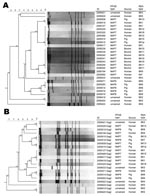
Figure 1. A). Dendrogram analysis of toxinotype V Clostridium difficile human and animal isolates using pulsed field gel electrophoresis (PFGE); SmaI restriction digest. Three animal-human isolate groups had indistinguishable PFGE patterns (2 NAP7...
All 8 recent human isolates had a 39-bp deletion in tcdC, and 6 (75%) of 8 were binary toxin positive. All 7 past human isolates also had a 39-bp deletion in tcdC, and 7 (100%) of 7 were binary toxin positive. Thirty-three toxinotype V animal isolates were obtained; they displayed a variety of PFGE patterns (Figure 1). All, however, were binary toxin positive and had a 39-bp deletion in tcdC. Three animal–human isolate groups had indistinguishable PFGE patterns (100% similarity) when digestion was performed with the SmaI enzyme. The first group contained 1 human isolate (REA subtype BK1) that was indistinguishable by PFGE (NAP7) from 1 porcine isolate (REA subtype BK13). The second group consisted of 5 human and 2 porcine isolates, all of which were designated NAP7 by PFGE, although REA demonstrated 4 different subtypes. The third group contained 1 human isolate and 2 porcine isolates, which were indistinguishable by PFGE and REA (NAP8 and BK6). The 9 isolates in groups 1 and 2 were only 80% similar when digestion was performed with EagI. However, the isolates in the third group were 95% similar, and 1 porcine isolate was indistinguishable (100% similarity) from 1 human isolate, even after digestion with EagI.
Figure 2
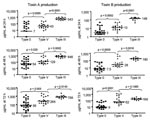
Figure 2. In vitro toxin production of toxinotype V Clostridium difficile isolates compared with epidemic toxinotype III and nonepidemic toxinotype 0 strains. Toxin A and Toxin B concentrations in micrograms per milliliter at...
Median toxin A and B production in the 21 toxinotype V isolates analyzed (7 bovine, 7 porcine, 7 of 8 recent human) was greater than that by nonepidemic toxinotype 0 isolates but less than that by epidemic toxinotype III isolates at all time points measured (Figure 2). The mean absorbance measurements at 600 nm, representing cell density, were measured at 24 h and 48 h and were not significantly different for toxinotype V isolates (1.56 and 1.06, respectively) than for toxinotype 0 isolates (1.77 and 1.39) or toxinotype III isolates (2.07 and 1.64).
Antimicrobial drug susceptibility testing was performed on 14 of 15 human and 16 of 33 animal toxinotype V isolates (Table 2). Resistance rates were similar overall in human and animal toxinotype V isolates except that more bovine isolates (88%) were susceptible to clindamycin than were porcine (0%, p<0.01) or human isolates (9%, p<0.01). All human and animal toxinotype V isolates (multiple strains by REA and PFGE) were susceptible to gatifloxacin and moxifloxacin, which differed markedly from human toxinotype III (BI/NAP1/027) and toxinotype 0 isolates (multiple strains by REA and PFGE).
In a review of recent and past isolates, we identified several human cases of CDAD caused by toxinotype V strains of C. difficile, which has been reported as a cause of epidemic disease in neonatal pigs and colonization in calves during the past decade (9,17,18). Moreover, different rates of occurrence in these temporally divergent populations suggest that toxinotype V may be an increasing cause of human CDAD, relative to other strains. The toxinotype V animal isolates included in our study have been previously identified as PCR ribotype 078, the most prevalent ribotype among calves and pigs, accounting for 94% (bovine) and 83% (swine) of isolates tested from multiple geographic regions (8). Food animal isolates we tested shared a high degree of similarity with human isolates, with 2 instances of animal–human isolate pairs appearing indistinguishable by REA or PFGE subtyping. In addition, all animal and human isolates displayed 39-bp deletions in tcdC, and most (45/47; 96%) were binary toxin positive.
Although C. difficile is recognized as a cause of disease in several animal species (19–22), little investigation has been conducted on the potential for interspecies transmission of C. difficile to humans. Previous studies have suggested the possibility of C. difficile transmission between humans and domestic pets (23,24), but no interspecies transmission has been documented, and few studies have examined the possible link between CDAD in food animals and humans. Identification of the same variant toxinotype strain as responsible for both human and animal disease in our study suggests at least 3 possible causes for human toxinotype V CDAD: 1) exposure of humans and animals to a common environmental source of C. difficile, 2) human disease caused by transmission by means of direct or indirect (e.g., through contaminated produce, water, or the environment) contact with infected live animals, and 3) human disease linked to consumption of products from food-producing animals. Both the genetic similarity of the human and animal isolates in our study and the apparent increasing importance of toxinotype V isolates in human CDAD after their emergence in animals may suggest foodborne or other forms of animal-to-human transmission.
In contrast to HCFA-CDAD, where patient-to-patient transmission of C. difficile is more likely, animal contact is a more plausible means of transmission for CA-CDAD. Our results suggest that toxinotype V C. difficile may be a relatively common cause of community-associated disease. Despite evidence that only 20% of all human CDAD cases are community-associated (25,26), 6 (46%) of 13 human toxinotype V cases in our study were identified as CA-CDAD. The high prevalence of CA-CDAD among toxinotype V cases we found is consistent with other studies that have identified variant toxinotypes more frequently in CA-CDAD than in HCFA-CDAD (27,28).
Toxinotype V strains may also be increasing as a cause of human CDAD since the emergence or recognition of epidemic toxinotype V disease in animals. In the past, reported frequencies of human strains with variant toxinotypes ranged from 6.4% to 13.4% of all C. difficile isolates collected (29–33), and toxinotype V strains contributed few cases to these frequency studies. However, a recent preliminary report from an Italian hospital indicated an upsurge in the proportion of binary toxin–positive C. difficile strains responsible for healthcare-associated disease in 2002 and 2003; most of these strains were toxinotype V (34). Toxinotype V appears to be an important cause of CDAD in food-producing pigs in parts of Europe, just as it is in North America (35).
The epidemiology of human CDAD has been affected by recent increases in the incidence and severity of disease. These changes have been largely attributed to the emergence of the BI/NAP1/027 C. difficile strain which, like the toxinotype V strains described here, is a toxin gene variant (i.e., toxinotype III) with an 18-bp deletion in tcdC (rather than the 39-bp deletion observed in toxinotype V strains) and with genes that encode binary toxin (4). In addition to this 18-bp deletion, and perhaps more importantly, BI/NAP1/027 has an upstream single nucleotide deletion at nucleotide position 117 (∆117), leading to a reading frameshift and early termination of protein translation (27).
Current literature suggests that this considerable truncation of TcdC may impair its negative regulatory function and contribute to the increased toxin production observed in BI/NAP1/027 strains (27,36). Molecular analysis of toxinotype V C. difficile has demonstrated a similarly truncated TcdC (61 aa compared with 65 aa in BI/NAP1/027 strains and 232 aa in wild-type TcdC) (15), which may imply hypervirulence for this strain as well. In contrast, isolates most commonly found in US hospitals before 2001 were toxinotype 0, had no polymorphisms in tcdC, and were binary toxin negative (37). Some of the increased virulence of BI/NAP1/027 may be due to its documented increased toxin A and B production in vitro (1). Although we did not find toxin production in toxinotype V isolates similar to BI/NAP1/027 levels, they did produce more toxin than nonepidemic toxinotype 0 isolates at all time points. Furthermore, the range of toxin A and B levels in our toxinotype V isolates was wide, and a minority produced toxin at similar or greater levels than BI/NAP1/027 strains.
This study is subject to the following limitations. First, the number of toxinotype V isolates examined was small and may not be wholly representative of this strain as it manifests in human or animal disease. Furthermore, recent isolates were collected from institutions that reported healthcare-associated outbreaks of CDAD, and clinical information describing patients from whom specimens were obtained may therefore overestimate disease severity. Moreover, since recent isolates were obtained from healthcare facilities that were experiencing CDAD outbreaks, they may represent a different population of patients than the past isolates, which were obtained from a variety of sources, some of which were ongoing clinical surveillance projects and some of which were outbreak investigations. The 2 source populations, however, can be considered reasonably similar in that both represent primarily hospitalized patients. If, however, the recent database contains a substantially greater proportion of outbreak-related isolates than the past collection, this would only strengthen the evidence for the recent emergence of toxinotype V CDAD. Since outbreaks of CDAD are largely associated with the epidemic, toxinotype III strain of C. difficile (4), relatively few toxinotype V isolates should be present in a recent database composed of outbreak-related isolates. The increased prevalence of toxinotype V in recent isolates compared with past ones may therefore represent an underestimate of the true prevalence of toxinotype V C. difficile.
Additionally, little is known about the types of C. difficile that cause disease in animals, which makes it impossible to determine whether the current toxinotype V strains are new or simply newly recognized. Finally, information about human cases is limited, particularly with respect to possible routes for community acquisition of disease; thus, evidence upon which to base conclusions regarding interspecies transmission is limited.
Although relatively common in animal CDAD, toxinotype V is currently an uncommon cause of human illness, which may occur more frequently among persons without traditional risk factors associated with CDAD, such as recent exposure to a healthcare setting. In vitro toxin production results from our limited sample suggest that toxinotype V strains have the potential to cause increased severity of human disease, although further studies are needed to corroborate this association. Although they share similar clinical features, evidence suggests that the predominant strains causing CDAD in humans and different animal species are distinct (8,38). Nonetheless, our finding of similarity between relatively widespread animal strains of C. difficile and strains responsible for occasional human disease raises the possibility of interspecies transmission. Further studies are needed to understand the etiology of CDAD caused by toxinotype V C. difficile and the mechanisms of transmission between animals and humans, including the role of the food supply.
Dr Jhung is a medical epidemiologist in the Division of Healthcare Quality Promotion at CDC. His primary interests include the epidemiology of healthcare-associated infections, outbreak investigations, infection control in resource-limited settings, and the public health response to disasters and emergencies.
References
- Warny M, Pepin J, Fang A, Killgore G, Thompson A, Brazier J, Toxin production by an emerging strain of Clostridium difficile associated with outbreaks of severe disease in North America and Europe. Lancet. 2005;366:1079–84. DOIPubMedGoogle Scholar
- Kuijper EJ, Coignard B, Tull P. Emergence of Clostridium difficile–associated disease in North America and Europe. Clin Microbiol Infect. 2006;12(Suppl 6):2–18. DOIPubMedGoogle Scholar
- Sunenshine RH, McDonald LC. Clostridium difficile–associated disease: new challenges from an established pathogen. Cleve Clin J Med. 2006;73:187–97.PubMedGoogle Scholar
- McDonald LC, Killgore GE, Thompson A, Owens RC Jr, Kazakova SV, Sambol SP, An epidemic, toxin gene-variant strain of Clostridium difficile. N Engl J Med. 2005;353:2433–41. DOIPubMedGoogle Scholar
- Songer JG, Post KW, Larson DJ, Jost BH, Glock RD. Enteric infection of neonatal swine with Clostridium difficile. Journal of Swine Health and Production. 2000;8:185–9.
- Songer JG, Post KW, Larson DJ, Jost BH, Glock RD. Clostridium difficile as a cause of porcine neonatal enteritis. Birmingham (AL): American Association of Veterinary Laboratory Diagnosticians; 2000.
- Post KW, Songer JG, Jost BH, Glock RD, Holtcamp A. The emergence of Clostridium difficile as a cause of porcine neonatal enteritis. Nashville (TN): American Association of Swine Veterinarians; 2001.
- Keel K, Brazier JS, Post KW, Weese S, Songer JG. Prevalence of PCR ribotypes among Clostridium difficile isolates from pigs, calves, and other species. J Clin Microbiol. 2007;45:1963–4. DOIPubMedGoogle Scholar
- Rodriguez-Palacios A, Staempfli HR, Duffield T, Peregrine AS, Trotz-Williams LA, Arroyo LG, Clostridium difficile PCR ribotypes in calves, Canada. Emerg Infect Dis. 2006;12:1730–6.PubMedGoogle Scholar
- Rodriguez-Palacios A, Staempfli HR, Duffield T, Weese JS. Clostridium difficile in retail ground meat, Canada. Emerg Infect Dis. 2007;13:485–7.PubMedGoogle Scholar
- McDonald LC, Coignard B, Dubberke E, Song X, Horan T, Kutty PK. Recommendations for surveillance of Clostridium difficile–associated disease. Infect Control Hosp Epidemiol. 2007;28:140–5. DOIPubMedGoogle Scholar
- Clabots CR, Johnson S, Bettin KM, Mathie PA, Mulligan ME, Schaberg DR, Development of a rapid and efficient restriction endonuclease analysis typing system for Clostridium difficile and correlation with other typing systems. J Clin Microbiol. 1993;31:1870–5.PubMedGoogle Scholar
- Klaassen CH, van Haren HA, Horrevorts AM. Molecular fingerprinting of Clostridium difficile isolates: pulsed-field gel electrophoresis versus amplified fragment length polymorphism. J Clin Microbiol. 2002;40:101–4. DOIPubMedGoogle Scholar
- Rupnik M, Avesani V, Janc M, von Eichel-Streiber C, Delmee M. A novel toxinotyping scheme and correlation of toxinotypes with serogroups of Clostridium difficile isolates. J Clin Microbiol. 1998;36:2240–7.PubMedGoogle Scholar
- Spigaglia P, Mastrantonio P. Molecular analysis of the pathogenicity locus and polymorphism in the putative negative regulator of toxin production (TcdC) among Clostridium difficile clinical isolates. J Clin Microbiol. 2002;40:3470–5. DOIPubMedGoogle Scholar
- National Committee for Clinical Laboratory Standards. Methods of antimicrobial susceptibility testing of anaerobic bacteria. 6th ed. Approved standard M11–A6. Villanova (PA): The Committee; 2004.
- Songer JG, Anderson MA. Clostridium difficile: an important pathogen of food animals. Anaerobe. 2006;12:1–4. DOIPubMedGoogle Scholar
- Songer JG. The emergence of Clostridium difficile as a pathogen of food animals. Anim Health Res Rev. 2004;5:321–6. DOIPubMedGoogle Scholar
- Frazier KS, Herron AJ, Hines ME II, Gaskin JM, Altman NH. Diagnosis of enteritis and enterotoxemia due to Clostridium difficile in captive ostriches (Struthio camelus). J Vet Diagn Invest. 1993;5:623–5.PubMedGoogle Scholar
- Marks SL, Kather EJ, Kass PH, Melli AC. Genotypic and phenotypic characterization of Clostridium perfringens and Clostridium difficile in diarrheic and healthy dogs. J Vet Intern Med. 2002;16:533–40. DOIPubMedGoogle Scholar
- Weese JS, Weese HE, Bourdeau TL, Staempfli HR. Suspected Clostridium difficile–associated diarrhea in two cats. J Am Vet Med Assoc. 2001;218:1436–9, 1421.
- Weese JS, Staempfli HR, Prescott JF. A prospective study of the roles of Clostridium difficile and enterotoxigenic Clostridium perfringens in equine diarrhoea. Equine Vet J. 2001;33:403–9. DOIPubMedGoogle Scholar
- O’Neill G, Adams JE, Bowman RA, Riley TV. A molecular characterization of Clostridium difficile isolates from humans, animals and their environments. Epidemiol Infect. 1993;111:257–64.PubMedGoogle Scholar
- Lefebvre SL, Arroyo LG, Weese JS. Epidemic Clostridium difficile strain in hospital visitation dog. Emerg Infect Dis. 2006;12:1036–7.PubMedGoogle Scholar
- Kutty PK, Benoit SR, Woods CW, Sena AC, Naggie S, Frederick J, Assessment of Clostridium difficile–associated disease surveillance definitions, North Carolina, 2005. Infect Control Hosp Epidemiol. 2008;29:197–202. DOIPubMedGoogle Scholar
- Noren T, Akerlund T, Back E, Sjoberg L, Persson I, Alriksson I, Molecular epidemiology of hospital-associated and community-acquired Clostridium difficile infection in a Swedish county. J Clin Microbiol. 2004;42:3635–43. DOIPubMedGoogle Scholar
- MacCannell DR, Louie TJ, Gregson DB, Laverdiere M, Labbe AC, Laing F, Molecular analysis of Clostridium difficile PCR ribotype 027 isolates from Eastern and Western Canada. J Clin Microbiol. 2006;44:2147–52. DOIPubMedGoogle Scholar
- Limbago B, Long CM, Thompson AD, Killgore GE, Hannett G, Havill N, Isolation and characterization of Clostridium difficile responsible for community-associated disease. International Meeting of the American Society for Microbiology; 2007 May 21–25; Toronto, Ontario, Canada.
- Barbut F, Gariazzo B, Bonne L, Lalande V, Burghoffer B, Luiuz R, Clinical features of Clostridium difficile–associated infections and molecular characterization of strains: results of a retrospective study, 2000–2004. Infect Control Hosp Epidemiol. 2007;28:131–9. DOIPubMedGoogle Scholar
- Geric B, Rupnik M, Gerding DN, Grabnar M, Johnson S. Distribution of Clostridium difficile variant toxinotypes and strains with binary toxin genes among clinical isolates in an American hospital. J Med Microbiol. 2004;53:887–94. DOIPubMedGoogle Scholar
- Rupnik M, Kato N, Grabnar M, Kato H. New types of toxin A-negative, toxin B-positive strains among Clostridium difficile isolates from Asia. J Clin Microbiol. 2003;41:1118–25. DOIPubMedGoogle Scholar
- Stubbs S, Rupnik M, Gibert M, Brazier J, Duerden B, Popoff M. Production of actin-specific ADP-ribosyltransferase (binary toxin) by strains of Clostridium difficile. FEMS Microbiol Lett. 2000;186:307–12. DOIPubMedGoogle Scholar
- Pituch H, Kreft D, Obuch-Woszczatynski P, Wultanska D, Meisel-Mikolajczyk F, Luczak M, Clonal spread of a Clostridium difficile strain with a complete set of toxin A, toxin B, and binary toxin genes among Polish patients with Clostridium difficile–associated diarrhea. J Clin Microbiol. 2005;43:472–5. DOIPubMedGoogle Scholar
- Buttrini M, Spigaglia P, Somenzi P, Zerbini L, Dettori G, Chezzi C, Epidemiology of Clostridium difficile strains with binary-toxin genes among clinical isolates in an Italian hospital. 2nd International Clostridium difficile Symposium; 2007 June 6–9; Maribor, Slovenia.
- Pirs T, Avbersek J, Ocepek M, Rupnik M. Isolation of Clostridium difficile from food animals in Slovenia. 2nd International Clostridium difficile Symposium; 2007 June 6–9; Maribor, Slovenia.
- Curry SR, Marsh JW, Muto CA, O’Leary MM, Pasculle AW, Harrison LH. tcdC genotypes associated with severe TcdC truncation in an epidemic clone and other strains of Clostridium difficile. J Clin Microbiol. 2007;45:215–21. DOIPubMedGoogle Scholar
- McFarland LV, Beneda HW, Clarridge JE, Raugi GJ. Implications of the changing face of Clostridium difficile disease for health care practitioners. Am J Infect Control. 2007;35:237–53. DOIPubMedGoogle Scholar
- Arroyo LG, Kruth SA, Willey BM, Staempfli HR, Low DE, Weese JS. PCR ribotyping of Clostridium difficile isolates originating from human and animal sources. J Med Microbiol. 2005;54:163–6. DOIPubMedGoogle Scholar
Figures
Tables
Cite This ArticleTable of Contents – Volume 14, Number 7—July 2008
| EID Search Options |
|---|
|
|
|
|
|
|
Please use the form below to submit correspondence to the authors or contact them at the following address:
Michael A. Jhung, Centers for Disease Control and Prevention, 1600 Clifton Rd NE, Mailstop A31, Atlanta, GA 30333, USA;
Top