Volume 14, Number 8—August 2008
Dispatch
Pathogenicity of Highly Pathogenic Avian Influenza Virus (H5N1) in Adult Mute Swans
Figure 2
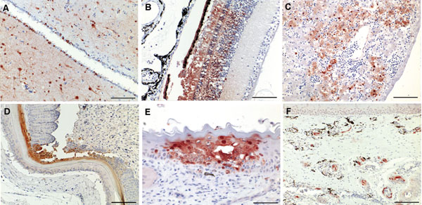
Figure 2. Immunohistochemical analysis for nucleoprotein of avian influenza virus. Tissue sections were stained by using the avidin-biotin-peroxidase complex method, 3-amino-9-ethylcarbazole (red), and hematoxylin (blue). A) Brain, cerebrum: numerous glial cells, neurons and ependymal cells stain positive for influenza virus antigen (scale bar = 200 μm). B) Eye, retina: cells of the pigmented epithelial layer, photoreceptor cells, and cells of the outer and inner nuclear layers are positive for the nucleoprotein of influenza virus (scale bar = 100 μm). C) Liver: subadjacent to the capsule there is hepatocyte degeneration and necrosis around a congested central vein (scale bar = 100 μm). D) Skin: keratinized layer of the feather follicular epithelium shows focal necrosis with intense nuclear and cytoplasmic immunostaining (scale bar =100 μm). E) Nasal cavity: focal intraepithelial necrosis of the mucocutaneous membrane associated with influenza virus infection (scale bar = 50 μm). F) Nasal concha: numerous submucosal arterioles and venules display strong endothelial staining, which partially extends into the media of the vessels (scale bar = 100 μm).